
UR Medicine

menu
UR Medicine / Dermatology / News


News

20242023202220212020

“Bubbles” Boost Search for Treatment to Aid Head and Neck Cancer Patients

Wednesday, October 25, 2017

Overby Photo

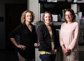
Catherine Ovitt, Danielle Benoit, and Lisa DeLouise

A scientific team at the University of Rochester is using innovative technology to discover preventative treatments for salivary gland radiation damage typical for head and neck cancer patients—and recently received a $3.8 million National Institutes of Health grant to support their investigation.

Cancer patients can lose salivary gland function during treatment for head and neck tumors. The irreversible damage, which prevents patients from producing saliva, often results in permanent dry mouth and makes it difficult to eat, speak, and swallow. The team will develop salivary gland tissues using a unique chip technology called "microbubbles," which are tiny spherical wells or bubbles that can hold cells.

The use of the microbubble platform is based on several years of salivary gland research, led by Catherine E. Ovitt, Ph.D., associate professor of Biomedical Genetics, a member of the UR Center for Oral Biology, and an expert in the repair and regeneration of salivary glands, and Danielle Benoit, Ph.D., associate professor of Biomedical Engineering and an expert in drug delivery systems and hydrogel platforms for tissue engineering approaches. Together with Lisa A. DeLouise, Ph.D., associate professor of Dermatology and Biomedical Engineering, who developed and received several patents for the microbubble concept, the scientists are working as co-principal investigators on the NIH project.

Their goal is to find drugs that could be given to patients prior to radiation treatment that would prevent damage to the glands.

"Dr. Ovitt and I have shown through years of investigation that being able to develop functional salivary gland tissue for testing is the key to solving this problem," Benoit said. "So, it's microbubbles to the rescue."

Expanding cells and tissue outside of the body is elusive. In this case the process involves taking salivary gland cells that have been removed from humans undergoing surgery, expanding the cells, and studying their reaction to various drugs.

A major problem, however, starts to occur as soon as the tissue is removed from the body and isolated: Cells immediately begin to lose their natural function. In the body, cells send signals and secrete proteins that are essential for their survival. In a culture plate in a laboratory, however, these signals and proteins are diluted and dispersed, making the cells no longer viable.

DeLouise's technology at first glance looks similar to a cell culture petri dish, a round piece of silicone about the size of the large cookie. But within the dish are an arrangement of thousands of tiny round "micro-wells," each one comprising a minuscule compartment for cell growth and tissue formation. The unique shape of each microbubble creates a niche that concentrates the cells, allowing them to proliferate and form salivary gland units.

The microbubbles come in different sizes, and the beauty of the technology is that scientists can grow cells in thousands of bubbles at one time. DeLouise can make dishes the size of a dime that include more than 5,000 microbubbles. In addition, Benoit's lab has produced hydrogel materials that can be placed inside each microbubble that further allow the cell to maintain its structure and function.

If the team can successfully grow human salivary gland cells in the microbubbles, they say, they will also be able to rapidly test thousands of existing Food and Drug Administration-approved drugs on the salivary tissue using the microbubble technology.

"Only one treatment is currently available for radioprotection but it comes with many side effects, so most patients discontinue it," Ovitt said. "There is a great need for additional ways to either cure or prevent this debilitating condition."

The team is collaborating with Shawn D. Newlands, M.D., Ph.D., M.B.A., chair of the Department of Otolaryngology and member of the Wilmot Cancer Institute's head and neck oncology team, to collect salivary tissue from consenting patients undergoing salivary gland surgery. Salivary gland cells are isolated from these tissues for seeding into microbubbles for the investigation. Additionally, Paul Dunman, Ph.D., associate professor of Microbiology and Immunology, will provide high-throughput drug-screening expertise during the second phase of the project, which is contingent upon successful development of the human gland chips.

Read More: “Bubbles” Boost Search for Treatment to Aid Head and Neck Cancer Patients

UR Medicine Dermatology Opens Brockport Office

Wednesday, August 16, 2017

UR Medicine Dermatology has opened an office in Brockport, led by dermatologist Kathryn E. Somers, M.D., assistant professor in the Department of Dermatology. Somers will be joined in September by a new faculty member, dermatologist/dermatopathologist Kathleen Mannava, M.D., who specializes in the pathology of skin conditions. Mannava, who was trained at SUNY Upstate Medical University, is relocating from Winston Salem, N.C. She will be an assistant professor in the Department of Dermatology.

The newest UR Medicine Dermatology location, in renovated space at 80 West Ave. , Suite 205, is part of the Strong West group of UR Medicine services. Strong West provides care to patients in western Monroe County and eastern Orleans and Genesee counties, including an Ambulatory Surgery Center, Imaging, Labs, UR Medicine Heart and Vascular, Primary Care, Orthopaedics, the Wilmot Cancer Institute, and Strong West Emergency.

Patient appointments will be available Tuesday through Friday. To refer a patient or book an appointment, please call (585) 275-7546.

Lisa A. Beck Invited to American Dermatological Association

Tuesday, August 1, 2017

Lisa A. Beck, M.D., Dean's Professor of Dermatology, was invited to membership in the American Dermatological Association (ADA), presented by Dermatology Chair Alice Pentland, M.D. ADA members are recognized leaders in Dermatology research, education, organizational medicine and patient care. ADA aims to further the advancement of all aspects of scientific endeavor in Dermatology. Membership is achieved through nomination and election based on meritorious contributions to the field of Medicine and Dermatology.

Poison Ivy: Prevention Takes Priority

Wednesday, June 21, 2017

Before heading out to dig in your garden, take steps to avoid a poison ivy encounter that might make you miserable. UR Medicine dermatologist Dr. Mary Gail Mercurio tells you what to look for and offers steps to take if poison ivy finds you before you find it.

poison ivy plant

Poison ivy, a toxic vine formally known as toxicodendron radicans, is the cause of a common—and often miserable—allergic skin reaction. Poison ivy is widespread in our region and its characteristic cluster of three almond-shaped leaves is familiar to many as is the adage: Leaves of three, let them be.

When exposed to the plant, people who are allergic can develop a red, itchy rash, sometimes with painful red bumps that often develop into fluid-filled blisters and tend to show up in lines or streaks corresponding with the areas of contact with the plant. The rash’s linear nature is one way to distinguish it from bug bites, also rampant in summertime. 

The precise culprit within poison ivy is urushiol, its oily sap. More than 75 percent of people are allergic to poison ivy, and the rash can begin anywhere from a few hours to a few days after exposure to the plant. Even if you haven’t had a problem with poison ivy, with repeated exposure, it’s possible to develop an allergy over time.

picture of poison ivy rash

The rash itself is not contagious. By the time it appears, the oil has either been washed off or absorbed into the skin. Touching the blister fluid cannot spread the rash. However, when the invisible oil gets on the hands and then other body parts are touched before the hands are washed, it is possible to unknowingly spread the rash widely, including to areas that came nowhere near the plant. Similarly, it is possible to get the rash from touching the oil on another person’s skin, a pet’s fur, or objects like gardening gloves or shoes that touched the plant and then contacted your skin before being washed. All of this happens long before a rash appears. Whenever there is a question of having been in contact with poison ivy, it’s wise to wash immediately with soap and water, and do the same for any clothing or objects to stop the spread.    

These tips may help you avoid the perils of poison ivy this summer:

  • Know it when you see it. Learn to recognize the plant so you can avoid it.
  • Block it. Consider using a barrier cream, which may reduce the risk of the plant’s oil penetrating your skin, in the event you are exposed to it.
  • Stop it in its tracks. If you think you’ve been exposed, do your best to wash the oil off your skin. 
  • Soothe it. If a rash does develop, work to keep it in check with an anti-itch or corticosteroid cream.
  • Get to know its counterparts. A variety of plants can cause rashes similar to poison ivy. Before spending time in the woods or garden, do a little online detective work to familiarize yourself with other culprits so you can enjoy being outdoors without worrying.
  • Seek help if it’s severe. In some instances, a poison ivy rash can be widespread or severe, involving the mouth, eyes, genitals or—worst of all—inhaled in smoke from a fire containing the leaves. In these cases, systemic corticosteroids and even hospitalization may be necessary. 

Mary Gail Mercurio, M.D.

Mary Gail Mercurio, M.D., is a professor of Dermatology and Obstetrics and Gynecology at URMC, caring for patients at UR Medicine’s Strong Memorial Hospital. She has special interest in skin and hair disorders in women.

The ABCs of SPF: Making Sense of Sunscreen Labels

Tuesday, June 13, 2017

Though some may find a suntan attractive, it's actually a sign of damage to skin cells—the kind of damage that may lead to skin cancer. And while it's the most common type of cancer, with proper sunscreen use, skin cancer is also the most preventable type of cancer.

UR Medicine dermatologist Dr. Sherrif Ibrahim offers some "ABCs" to help you learn what to look for when shopping for sunscreen and how to decipher some of the labeling language prompted by new FDA regulations.

father applying sunscreen to his son

A: All-ray protection: Make sure your sunscreen is labeled as "broad spectrum." That means it protects against both UV-A and UV-B rays, both of which can lead to skin cancer.

B: Be SPF wise: The sun protection factor, or SPF, should be at least 30 but doesn't need to be higher than 50. According to the FDA's new labeling, anything under SPF 15 does not adequately prevent skin cancer. The higher the SPF, the better; however, once you get up to 50 SPF, about 98 percent of UV rays are filtered out. New labeling will eliminate SPF values beyond 50.

C: Caution when wet: Sunscreen should no longer be labeled as "waterproof" but will instead say "water-resistant" and will include the maximum time it will be effective in water, usually 40 or 80 minutes. This language is more accurate than previous language because no sunscreen is truly waterproof and it needs to be reapplied frequently, which is reiterated by the timing given on the label. It's also worth mentioning that "sport" sunscreen effectively protects you even when sweaty as long as it's applied prior to sweating so that it can bond to the skin's surface.

D: Do it all day long: No matter what brand you buy, all sunscreens need to be reapplied throughout the day to be effective. It's smart to reapply an amount that would fill a shot glass to the exposed areas of the body (face, arms, and legs) at least every couple of hours. For those who have trouble remembering to reapply when you're out in the sun, new smart phone apps can alert you when it's time to slather on more. There are also devices you can wear that can tell you when you've reached the maximum amount of UV exposure you should receive that day—so you know when it's time to go inside. Usually UV rays are strongest between 11 a.m. and 2 p.m. so it's best to avoid being outside during that time if possible.

In Rochester and around upstate New York, every day of sunlight is a gift, but for unprotected skin, sun causes more harm than good. Anything you can do to help protect your skin from burns and tans will reduce your chances of getting skin cancer not while keeping your skin looking younger for longer.

Enjoy your summer safely this year!

Sherrif Ibrahim MD

Sherrif Ibrahim, M.D., Ph.D., is a dermatologist with UR Medicine's Wilmot Cancer Institute. His focus is on the diagnosis, treatment, and prevention of skin cancer.

University start-ups highlighted in national innovation report

Thursday, April 27, 2017

Two University start-up companies are among those singled out in a new report from the Science Coalition. The report, “American-Made Innovation Sparking Economic Growth,” identifies 102 companies that trace their roots to federally funded university research.

“The innovation that drives economic growth in the U.S. is based, in large part, on the scientific discoveries made in research universities and funded by the federal government,” said Rob Clark, University provost and senior vice president for research. “As a nation, it is imperative that we continue to support the fundamental science that leads to new technologies and improves lives.”

One of the companies is Adarza Biosystems was founded in 2008 by University of Rochester researchers Ben Miller and Christopher Striemer. The company, which has operations in Rochester and St. Louis, is developing a microchip device that can be used to detect proteins in biological samples such as blood. The technology is being developed for both the lab and the clinic and could be used to improve cancer diagnostics and allergy testing, drug and vaccine development, and research on infectious diseases. The technology was developed with the support of a $1 million grant from the NIH.

Read More: University start-ups highlighted in national innovation report

FDA Approves New Eczema Drug Tested at URMC

Thursday, March 30, 2017

People who suffer from eczema have new hope for relief with the U.S. Food and Drug Administration’s approval of a “game changing” medication tested at the University of Rochester Medical Center (URMC). The new drug, Dupilumab, is the first treatment to show significant, lasting results for adults whose lives are disrupted by moderate to severe eczema or atopic dermatitis (AD).

AD is the most common skin disease in the world, impacting 7 to 9 percent of adults. People with AD suffer from severely dry skin, red lesions that may crust or ooze, skin thickening and intense itching, which may lead to skin wounds, infections, sleep disturbance and depression.

“This is a watershed moment for the treatment of patients with AD, some of whom have been suffering for decades with intractable itch and extensive skin disease,” said Lisa A. Beck, M.D., Dean’s Professor of Dermatology at URMC and a leading researcher in clinical trials of the new drug. “Until now, we’ve had nothing new to offer patients with this level of disease. This is a real game-changer for them.”

At URMC, Beck led a three-year trial of the drug to test its efficacy and safety. In trials, Dupilumab rapidly and significantly improved the skin appearance and the severity of itch in patients. In addition, patients had little or no side effects from the medication.

Read More: FDA Approves New Eczema Drug Tested at URMC

Partnership Will Help Create “Eyes and Ears” for Photonics Technologies

Wednesday, March 29, 2017

A new Defense Department project will help researchers develop sensors that can be employed by photonics-based systems. The sensors – which could be used in a wide range of applications, such as environmental monitoring, disease diagnosis, detection of chemical and biological weapons, and to ensure food safety – represent a key component of the AIM Photonics initiative.

The $900,000 U.S. Department of Defense (DOD) project, along with an additional $1.41 million in matching funds from AIM Photonics industrial members, will support a consortium of partners led by the University of Rochester that includes the U.S. Army, U.S. Navy Research Lab, Ortho-Clinical Diagnostics, Analog Photonics, the University of Tulsa, Phoenix, the University of California-Santa Barbara, and OndaVia.

“When you consider the impact these sensors will have in immediately diagnosing illness, rapidly detecting foodborne pathogens, instantly assessing water and air quality, and ensuring the security of our citizens, you then realize the significance of AIM Photonics and how the technology we are developing is nothing short of a revolution,” said John Maggiore, chairman of the New York State Photonics Board of Officers.

“Sensors represent the interface between the real world and data,” said Ben Miller, Ph.D., the principal investigator of the project. “Developing a universal set of protocols to design, manufacture, modify, and integrate sensors into photonics systems will not only advance this technology, but also present a tremendous economic opportunity—integrated photonics sensors represent a large and rapidly growing market, potentially reaching more than $15 billion globally by 2020.”

Read More: Partnership Will Help Create “Eyes and Ears” for Photonics Technologies

DeLouise Gives Rochester Science Cafe Talk

Tuesday, March 28, 2017

Dermatology associate professor, Lisa DeLouise, MD, MPH, gave a 'Science Cafe' talk last night entitled, "Skin and the Environment: Things You Should But Probably Don't Know".

The DeLouise Bio Nanomaterials Group manipulates materials on the micro, nano and molecular level scales to design biomedical devices for diagnostic, therapeutic and investigative studies of skin. Our multidisciplinary approach integrates experimental research and simulation to design devices that exploit the novel optical, morphological and surface chemical properties of nanostructured materials. Key focus areas include optical biosensing, microfluidic cell sorting and cell culture and nanoparticle skin penetration, skin imaging, translocation and targeted delivery.

Science Cafes are interactive events involving face-to-face conversations with leading scientists about relevant current topics. This one meets on the fourth Tuesday of the month in the Community Room on the second Floor of the Pittsford Plaza Barnes and Noble.

Read More: DeLouise Gives Rochester Science Cafe Talk

Helping Doctors Diagnose and Treat Psoriatic Arthritis

Monday, March 13, 2017

Doctor examining an elderly woman's jointsDespite decades of research on psoriatic arthritis (PsA), diagnosing this debilitating inflammatory joint and skin disease is often delayed. Because the disease varies so much from patient to patient, researchers have not been able to hone in on a clear set of clinical criteria to diagnose it. URMC researchers provide some guidance to help doctors recognize signs of PsA and treat the disease earlier in a review recently published in the New England Journal of Medicine.

PsA is essentially a grab bag of mixed and matched symptoms. While PsA always occurs alongside psoriasis, an immune skin disorder characterized by painful, red, scaly patches of skin, other symptoms are less reliable. PsA patients may have inflamed, swollen fingers or toes referred to as "sausage fingers", or may experience any combination of inflammation in tendons, ligaments, the lower spine, and/or peripheral joints (any joint other than the spine). One distinguishing characteristic of PsA is its penchant for destruction of cartilage and bone and harmful creation of new bone, which is unparalleled by other forms of arthritis.

These symptoms can also come and go -- flaring up in response to emotional or immune stress. Patients' daily living can be significantly impacted during flare ups, making it hard for them to maintain productivity and often causing them to miss work.

Christopher Ritchlin, M.D., M.P.H., professor and chief of Allergy, Immunology, and Rheumatology at the University of Rochester Medical Center and author the review, has studied PsA for over 25 years and has treated many PsA patients in the clinic. He and review co-authors Dafna Gladman, M.D., of Toronto Western Hospital and Robert Colbert, M.D., Ph.D., of the National Institute of Arthritis and Musculoskeletal and Skin Diseases, offer their collective expertise on testing for PsA and differentiating it from similar forms of arthritis.

The group recommends assessing a litany of joints for tenderness and soft swelling due to inflammation, occurring in a unique "ray pattern"; each joint of a single finger may be affected, while neighboring fingers are entirely spared. Physicians should also evaluate spinal range of motion and back pain as well as the severity of psoriasis, indicated by the amount of skin that appears inflamed and signs of nail pitting or separation from the nail bed.

Because PsA varies so much from patient to patient, the review authors suggest physicians tailor treatment strategies to specific, dominant features of the disease in each patient. Non-steroidal anti-inflammatory drugs (NSAID) may be sufficient for some, while others may need one of the many disease modifying anti-rheumatic drugs (DMARDS) now available.

All PsA patients, however, can benefit from lifestyle modifications that keep inflammation at bay. Quitting smoking, maintaining a healthy body weight, staying physically active, managing stress, and protecting joints are some vital changes that PsA patients can make to help manage their disease.

For more advice on diagnosing and treating PsA, read the full review article.

Read More: Helping Doctors Diagnose and Treat Psoriatic Arthritis