News
20242023202220212020
Congratulations Dr. Matthew H. Ingalls!
Saturday, December 4, 2021
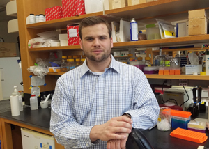 This week, on Friday December 3rd, GDSC graduate student Matthew H. Ingalls defended his doctoral thesis titled, ‘Acinar Cell Dynamics in Salivary Gland Homeostasis and Recovery from Injury’. During his time at the University of Rochester, Matt studied the role of cellular plasticity in tissue regeneration, salivary gland regeneration mouse genetic models, and tissue engineering. Matt did his thesis research under the mentorship of his advisor Dr. Catherine Ovitt, Ph.D. We wish Matt the best as he embarks as a Postdoctoral Researcher in the Batista Lab at Washington University of Medicine in St. Louis, Missouri.
Matt’s thesis abstract can be read below.
Congratulations Matt!
Abstract: Acinar Cell Dynamics in Salivary Gland Homeostasis and Recovery from Injury By Matthew Ingalls
Salivary gland dysfunction, resulting from the loss of secretory acinar cells, is caused by radiation therapy, duct obstruction, or the autoimmune disease Sjögren’s syndrome. To date, there is no effective therapeutic means to restore acinar cells and there has been much debate within the field as to which endogenous cell population should be the focus for such endeavors. A growing body of research indicates that acinar cells themselves may serve as a pool for maintenance of the acinar cell population without a bona-fide stem cell. Furthermore, the literature suggests that acinar cells utilize a mechanism involving cellular plasticity to contribute to recovery from acute injury.
 Research presented in this thesis investigated the source of acinar cell maintenance in human salivary glands using histology and explant tissue culture experiments. Like mouse acinar cells, it was found that endogenous human salivary gland acinar cells are mitotically active and directly support the acinar cell population under non-injury conditions. This study also demonstrates that the seemingly homogenous acinar cell population is made up of two separate populations, expressing all characteristic acinar cell markers that can be distinguished by a mononucleated or binucleated state. Finally, these studies set out to determine a clear role for cellular plasticity in salivary gland acinar cell injury response and recovery. Using an inducible acinar cell lineage tracing mouse model, it was found that shortly after duct ligation injury, acinar cells undergo plasticity, namely an acinar-to-ductal transition. Upon injury, the acinar lineage loses expression of acinar-specific markers and gains expression of the duct marker, cytokeratin-7. Concomitantly, a reduction in mTOR activity, cessation of proliferation, a brief activation of genes involved in the secretion of pro-inflammatory cytokines, and loss of characteristic cell-cell contacts is observed. In recovery, the acinar cell lineage regains mTOR activity, loses duct cell characteristics, expresses acinar cell markers, and resumes proliferation. Given the changes which occur in the acinar cell population during duct ligation, we speculate that the process of cellular plasticity in salivary gland involves an evolutionarily-conserved mechanism of tissue regeneration, called paligenosis. Previous work has shown that similar changes occur in the acinar cell lineage in response to radiation. However, unlike the response to duct ligation, months following radiation injury, mTOR suppression is sustained, pathways involved in the secretion of pro-inflammatory cytokines are not resolved, and the secretory acinar cell population does not recover to the same degree.
Collectively, these data support the notion that cellular plasticity in salivary glands occurs via a mechanism similar to paligenosis and suggest that it is required for recovery of functional salivary gland acinar cells. Future studies to understand differences between the injury response to duct ligation and radiation injury will determine whether perturbing mTOR or other targets involved in paligenosis will allow the endogenous acinar cell population to serve as a source for regeneration in a clinical setting.
Congratulations Dr. Jayme Olsen!
Monday, November 29, 2021
 On Monday November 29th, GDSC graduate student Jayme Olsen defended her thesis titled ‘Bmi-1 Regulation of Human Erythroid Ex Vivo Self-Renewal’. Jayme pursued her graduate research under the mentorship of Dr. James Palis, MD.
Jayme’s thesis abstract can be read below.
Congratulations Jayme!
Abstract: Bmi-1 Regulation of Human Erythroid Ex Vivo Self-Renewal By Jayme L. Olsen
 Human erythroid progenitors derived from adult sources are capable of restricted ex vivo self-renewal when cultured with erythropoietin, stem cell factor, and dexamethasone prior to their terminal maturation. This limited capacity for ex vivo self-renewal is a major obstacle in generating sufficient numbers of red blood cells to be useful for clinical and research applications. We previously discovered that erythroblasts derived from the murine embryo have a unique ability to extensively self-renew ex vivo, and that Bmi-1, a member of the polycomb repressive complex 1, is both necessary and sufficient to drive the extensive self-renewal of adult murine marrow-derived erythroblasts. Here we tested the hypothesis that Bmi-1 regulates the ex vivo self-renewal of human erythroid cells. Lentiviral overexpression of Bmi-1 increased the proliferative capacity of adult human peripheral blood mononuclear cell-derived erythroblasts over ten billion-fold. Bmi-1-induced self-renewing erythroblasts (iSREs) retained an immature erythroid morphology and cell surface phenotype and remained dependent on cytokines throughout prolonged culture. Analysis of gene expression data revealed that an increase in Bmi-1 resulted in a downregulation of multiple negative regulators of the cell cycle. Both chemical inhibition of Bmi-1 and lentiviral knockdown of Bmi-1 resulted in a massive reduction in cycling cells and an increase in cell death which led to a collapse of the culture. These data indicated that Bmi-1 is both necessary and sufficient to increase the ex vivo self-renewal capacity of human erythroid cells. Bmi-1 overexpression did not interfere with the ability of iSREs to mature in vitro based on morphology and cell surface phenotype. iSRE-derived reticulocytes successfully underwent serologic testing of multiple antigens using the gel card assay. Overall, increasing the ex vivo self-renewal capacity of human erythroid cells ultimately paves the way for the generation of sufficient numbers of cultured RBCs for the establishment of robust in vitro models of RBC-intrinsic disorders, for improved blood typing and transfusion therapy.
BGG Graduate Student Jamie Burchett Passes his Qualifying Exam!
Tuesday, October 5, 2021
 BGG Graduate Student Jamie Burchett, from the lab of Dr. Brian Altman, passed his Qualifying Exam. Jamie's thesis proposal is titled "Determining the role of REV-ERBs in cancer downstream of MYC".
Abstract: The MYC family of genes are a set of transcription factors that control several core biological processes important to cancer cell initiation and growth, such as cell cycle progression, metabolism, and biomass accumulation. They are amplified, translocated, or otherwise overexpressed in many types of cancers, and their disruption is often linked to poorer prognosis. Two of MYC's known downstream targets are NR1D1 and NR1D2 (the REVERB proteins), nuclear hormone receptors that act as transcription repressors with a variety of targets. REVERB α and β repress BMAL1 (ARNTL) production, a core clock protein which, together with CLOCK, is responsible for the regulation and oscillation of pathways and processes known as the molecular clock. BMAL1 and its binding partners are important regulators on both a cellular and whole-body level, and are responsible for a wide range of cellular processes such as metabolic rates, cell growth and proliferation, and apoptosis. This repression of BMAL1 by MYC leads to the overall disruption of the clock and its target pathways, but the mechanisms through which these REVERB molecules, BMAL suppression, and clock disruption support altered metabolism and tumorigenesis are still not well understood. While the interactions between MYC, REVERB, and the core clock proteins are established, it is important to fully understand what REVERBs are doing downstream of MYC, both with their primary targets and downstream of their effector pathways due to the potential of REVERB's being therapeutic targets in cancer. To ask these questions, we will study non-small cell lung cancer (NSCLC), since it has previously been characterized to be affected by circadian disruption and have increased MYC levels. We hypothesize that the induction of REVERB molecules by MYC has pro-tumorigenic effects through both direct effects on metabolism and cell growth and downstream effects through BMAL1 inhibition. We will be addressing this hypothesis through three different aims. In the first Aim, we will analyze the effects of REVERB on cell growth and tumorigenesis in cancer cell lines through colony formation, ability to grow at low concentrations, and tumor initiation. In our second Aim, we will study how REVERB affects metabolism of MYC-driven cells by studying respiration, glycolysis, and lipogenesis rates followed by nutrient deprivation to verify any revealed sensitivities. Finally, we will utilize bioinformatic analysis of publicly available human tumor data to determine the effect of MYC amplification on circadian rhythms, their gene expression, and identifying common pathways altered in varying tumor types.
GDSC Graduate Student Ludia J. Pack Passes her Qualifying Exam!
Tuesday, October 5, 2021
 GDSC Graduate Student Ludia Pack, passed her Qualifying Exam. Last year, Ludia joined the laboratory of Dr. Mark Noble. Ludia’s thesis research focuses on restoring Redox/Fyn/c-Cbl (RFC) pathway function as a therapeutic target in non-small cell lung cancers and ovarian cancers. Ludia's thesis proposal abstract can be read below. Congratulations Ludia!
Abstract
Non-small cell lung cancers (NSCLCs) are the most lethal subtype of lung cancers, which are the number one cause of cancer-related death worldwide. Despite the development of numerous treatments, there is still no cure for NSCLCs. There are two major subcategories of NSCLCs: lung adenocarcinomas (LUADs) and lung squamous cell carcinomas (LUSCs). Although the molecular landscape of NSCLC is very heterogeneous, there are actionable pathways of interest that have led to the development of targeted therapies such as tyrosine kinase inhibitors. Similarly, if the tumor expresses high levels of programmed-death ligand 1 (PD-L1), immune checkpoint inhibitors are included in the therapeutic regimen. However, the current obstacles to the success of existing therapies result from the development of primary, adaptive, or secondary resistance, highlighting the need for more research and development of additional therapies. In addition, no targeted therapies exist for LUSCs.
Many of the targets of interest in both LUADs and LUSCs are targets that can be modulated by the E3 ubiquitin ligase known as Casitas B-lineage lymphoma (c-Cbl). C-Cbl is an important tumor suppressor, due to its capability of negatively regulating mitogenic pathways via ubiquitination of receptor tyrosine kinases as well as non-receptor tyrosine kinases. C-Cbl has also been shown to play a role in decreasing levels of PD-L1. Previous studies in our lab have discovered that c-Cbl can become super-activated via the Redox/Fyn/c-Cbl (RFC) pathway. The RFC pathway becomes activated by increases in intracellular oxidation, which leads to the phosphorylation of Fyn kinase, subsequently causing the phosphorylation and super-activation of c-Cbl, which leads to enhanced degradation of its targets. The lab found that c-Cbl can become inhibited by the rho-guanine nucleotide exchange factor ßpix, and that we can reverse this inhibition via an FDA approved antidepressant called CRA01 (for c-Cbl restorative agent -1).
The pathways of therapeutic interest in NSCLCs may be targetable by harnessing the RFC pathway. My proposal aims to determine if c-Cbl is inhibited in NSCLCs, whether CRA01 can restore c-Cbl function, and if activation of the RFC pathway is a viable strategy for treating cancer growth in vitro and in vivo. The second part of my proposal aims to determine if super-activation of c-Cbl leads to enhanced degradation of PD-L1, and whether this increased degradation leads to increased immune cell infiltration in immuno-competent mice, allowing for the development of a treatment strategy that tackles multiple aspects of NSCLC tumor biology.
GDSC Student, Adrián Moisés Molina Vargas, accepts award on behalf of Alliance for Diversity in Science & Engineering (ADSE) Student Group
Monday, September 20, 2021
Today the University of Rochester recognized the Alliance for Diversity in Science & Engineering (ADSE) student group with the Trainee Diversity Award for their support of underrepresented groups pursuing STEM education. GDSC Student, Adrián Moisés Molina Vargas accepted the ADSE's award with a group of fellow ADSE members. Adrián serves as the ADSE's Social Media Chair and GSS Representative. He pursues his doctoral research in the lab of Dr. Mitchell R. O'Connell, Ph.D.
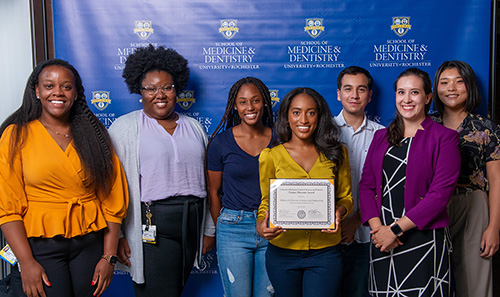
Congratulations to all the members of the ADSE!
To learn more about the ASDE and their mission, you can go their websites below:
GDSC Students Katherine (Katie) Padilla and Bachelard (Bache) Dieujuste Recognized at Convocation Award Ceremony
Monday, September 20, 2021
Earlier today two first-year GDSC students were presented with awards at the UR's Annual Opening Convocation Ceremony.
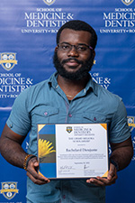
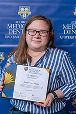
Katherine (Katie) Padilla was awarded a Provost’s Fellowship. This award is designed to help us recruit talented individuals who will broaden the diversity of those pursuing Ph.D. degrees at the University of Rochester. Bachelard (Bache) Dieujuste was awarded a Meliora Scholarship. This new award recognizes incoming Ph.D. students that demonstrate strength of character and exceptional promise for success.
Katie and Bache, along with our three other first-year students, Riti Kamath, Kira Taylor, and Jingyi Wu, are off to a great start and we’re delighted to have all of them join in the GDSC program.
GDSC Graduate Student Brandon Park Passes his Qualifying Exam!
Thursday, August 26, 2021
 Completing his 2nd year on a high note, GDSC Graduate Student Brandon Park, passed his Qualifying Exam. Brandon plans to focus his research on Nucleic Acid Biology, Epigenetics, and Development. His thesis proposal is titled “Using New Technologies to Investigate the Role of H2A.Z in Epigenetic Inheritance”. Brandon is pursuing his doctoral research under a co-mentorship with Dr. Mitch O’Connell, and Dr. Patrick J. Murphy.
Congratulations Brandon!
Brandon Park’s thesis proposal may be read below.
Abstract: 5-methylcytosine (5mC) is a major form of DNA methylation that is often observed at the promoters and enhancers of repressed genes, but the mechanisms by which 5mC patterns are maintained in developing primordial germ cells (PGCs)is currently unknown. Nucleosomes that contain the histone variant H2A.Z function as “placeholders” in early embryos to deter DNA methylation following fertilization, and are thus located at hypomethylated regions, such as promoters for genes involved in early development. Therefore, I hypothesize that H2A.Z maintains this function during PGC development, allowing the DNA methylation patterns of stem cells to be maintained in PGCs. This mechanism would facilitate the trans-generational maintenance of 5mC patterns and might enable adaptive evolution if changes to these 5mC patterns are inherited. To date, technical limitations of Chromatin Immuno-Precipitation (ChIP) have made it difficult to assess the localization of chromatin modifications throughout PGC development and in mature oocytes. CUT&TAG is a recently developed alternative to ChIP-Seq that provides significantly improved signal to noise and can be done on just a few thousand cells. For this reason, I will use CUT&TAG to investigateH2A.Zpatterns in PGCs that have been purified via Fluorescence Assisted Cell Sorting (FACS), as well as whole WT sperm, oocytes, and early embryos. I will bioinformatically analyze the resulting H2A.Z landscapes to define regions where H2A.Z patterns change or are maintained in different cell types. These experiments will create a reference point for defining H2A.Z landscapes, and test our hypothesis that H2A.Z patterns are maintained throughout germ cell development. Functional studies of PGCs have also been limited by the availability of genetic tools in zebrafish, as well as difficulties generating tissue specific knockdown effects. Here I will create a tissue-specific CRISPR system to knockdown H2A.Zat various time points during PGC development using a heat shock inducible promoter to control gRNA expression. I will investigate the effects of these knockdowns by doing CUT&TAG for H2A.Z, locus specific bisulfite sequencing (through PCR), and monitoring for phenotypic changes in the mature germ cells and next generation embryos. Together, these experiments will combine CRISPR and CUT&TAG methods to establish several epigenomic profiles for various factors involved in regulating gene expression in developing zebrafish embryos and PGCs. These studies will also provide new insight into the role of H2A.Z in 5mCmaintenance and transgenerational epigenetic inheritance.
Appetite for survival: Brain signal alerts roundworms to changing food supply
Wednesday, August 25, 2021
Microscopic roundworms may hold the key to understanding what is happening in the brain when the instinct of an animal changes in order to survive. In a newly published paper in the journal Current Biology University of Rochester Medical Center researchers found that a signaling system in the brain changes to redirect the behavior of an animal when their survival is at risk because there is not enough food.
The experiments were conducted in C. elegans – a microscopic roundworm that has been used by scientists for decades to understand the basic organization and function of the central nervous system and how it impacts behavior. Researchers found C. elegans hermaphrodites (the equivalent of females in this species) produce a pheromone that allows worms to monitor how crowded their environment is and how much food there is to go around. When food becomes scarce the aversion circuit is trigged in the animal and it becomes repelled by the pheromone.
“The key thing we identified is a molecular mechanism whereby an instinctive response can be suppressed under particular environmental conditions, namely, abundant food,” said Douglas Portman, Ph.D., lead author of the study and professor of Biomedical Genetics. “Adaptively it makes sense that an animal’s instinctive response would have this kind of flexibility.”
This underlying repulsive mechanism to the pheromone is present in both hermaphrodites and males, but researchers found that in males, the mechanism is overridden by another circuit that causes males to be attracted to the pheromone. A subtlety that could provide an understanding of how the neural circuits work that cause this change in behavior.
Understanding how basic decision-making mechanisms work gives insight into the inner workings of a more complex brain. “These findings lend important insight into the mechanisms by which animals detect and integrate multiple sensory cues to make adaptive behavioral decisions. Understanding how things like this work at the molecular level, provides a framework for understanding how much more complex brains work, and how genetic and environmental insults can ‘break’ things and lead to behavioral and psychiatric disorders.”
Postdoctoral fellow Jintao Luo, Ph.D., with the University of Rochester, co-authored this study. The research was supported with funding from the National Institute of General Medical Sciences.
Read More: Appetite for survival: Brain signal alerts roundworms to changing food supplyGDSC Graduate Student Emily Berry Passes her Qualifying Exam!
Tuesday, July 27, 2021
 GDSC Graduate Student, Emily Berry successfully passed her qualifying exam. Last summer, Emily joined the lab of Stephano Spano Mello, Ph.D. Emily’s doctoral research project is titled “Defining the mechanisms underlying Neat1’sfunction in preserving pancreas cell identity”. Emily’s research abstract is sampled below.
Congratulations Emily!
Abstract: Intraductal papillary mucinous neoplasms (IPMNs) are benign pancreatic lesions whose diagnosis has been increasing steadily over the last few years. IPMNs are slow-growing lesions originating from pancreatic duct cells. IPMNs can sometimes progress into invasive pancreatic ductal adenocarcinoma (PDAC), a deadly disease with poor patient survival. Therefore, IPMNs represent an opportunity to diagnose and treat a deadly disease before it advances to metastatic PDAC. Downregulation or mutation of protein subunits in the SWItch/Sucrose Nonfermentable (SWI/SNF) chromatin remodeling complex has been linked to IPMN formation, however the exact molecular mechanisms behind IPMN formation and malignant development are poorly understood. We recently showed that thep53-induciblelincRNA Nuclear Enriched Abundant Transcript 1 (Neat1) acts as a tumor suppressor in PDAC. We observed increased IPMN lesions in mouse models of PDAC lacking Neat1, similar to what was observed in SWI/SNF deficient mice. Additionally, a direct interaction between Neat1 and the SWI/SNF catalytic subunit BRG1 has been reported in some cell types. Based on these observations, we postulated that Neat1and SWI/SNF could be acting on the same path to suppress IPMN formation. My preliminary data show that loss ofNeat1leads to genome-wide changes in chromatin accessibility and SWI/SNF localization in both mouse embryonic fibroblast (MEFs) and in pancreatic lesion cells from mouse models of PDAC. I therefore hypothesize that Neat1 restricts IPMN formation by impacting SWI/SNF-dependent chromatin remodeling. To test this hypothesis, I propose to; 1) define the interaction between Neat1 and SWI/SNF in the pancreas, 2) determine how DNA accessibility and SWI/SNF localization on chromatin are impacted by loss of Neat1,and 3) determine which genes are differentially expressed upon the loss of Neat1, and how these genes may contribute to IPMN formation and malignancy. These experiments will illuminate the role of Neat1 and SWI/SNF in the formation of IPMN lesions.
Congratulations Xiaolu!
Thursday, June 17, 2021
 We wish a double congratulations to GDSC Graduate Student Xiaolu Wei. At the recent 32nd Genetics Day Symposium, her poster entry, titled "Intragenomic Conflict in Drosophila: Satellite DNA and Meiotic Drive" earned, not one, but two awards! First, symposium judges selected Xiaolu's poster, to be one of the six Short Talk Competition finalists. Then on Genetics Day, after the six finalists presented their Short Talks live, Xiaolu Wei's poster was voted the Fan Favorite among all Genetics Day attendees. Xiaolu's research in the Larracuente Lab combines genomic, and molecular techniques to discern how large blocks of repetitive DNA (satellites) are regulated in the germline.
We hope this was our last virtual Genetics Day for some time, and that at next year's Genetics Day, we may congratulate our Symposium finalists and fan favorites in person.