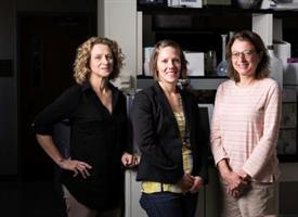
Catherine Ovitt, Danielle Benoit, and Lisa DeLouise
A scientific team at the University of Rochester is using innovative technology to discover preventative treatments for salivary gland radiation damage typical for head and neck cancer patients—and recently received a $3.8 million National Institutes of Health grant to support their investigation.
Cancer patients can lose salivary gland function during treatment for head and neck tumors. The irreversible damage, which prevents patients from producing saliva, often results in permanent dry mouth and makes it difficult to eat, speak, and swallow. The team will develop salivary gland tissues using a unique chip technology called "microbubbles," which are tiny spherical wells or bubbles that can hold cells.
The use of the microbubble platform is based on several years of salivary gland research, led by Catherine E. Ovitt, Ph.D., associate professor of Biomedical Genetics, a member of the UR Center for Oral Biology, and an expert in the repair and regeneration of salivary glands, and Danielle Benoit, Ph.D., associate professor of Biomedical Engineering and an expert in drug delivery systems and hydrogel platforms for tissue engineering approaches. Together with Lisa A. DeLouise, Ph.D., associate professor of Dermatology and Biomedical Engineering, who developed and received several patents for the microbubble concept, the scientists are working as co-principal investigators on the NIH project.
Their goal is to find drugs that could be given to patients prior to radiation treatment that would prevent damage to the glands.
"Dr. Ovitt and I have shown through years of investigation that being able to develop functional salivary gland tissue for testing is the key to solving this problem," Benoit said. "So, it's microbubbles to the rescue."
Expanding cells and tissue outside of the body is elusive. In this case the process involves taking salivary gland cells that have been removed from humans undergoing surgery, expanding the cells, and studying their reaction to various drugs.
A major problem, however, starts to occur as soon as the tissue is removed from the body and isolated: Cells immediately begin to lose their natural function. In the body, cells send signals and secrete proteins that are essential for their survival. In a culture plate in a laboratory, however, these signals and proteins are diluted and dispersed, making the cells no longer viable.
DeLouise's technology at first glance looks similar to a cell culture petri dish, a round piece of silicone about the size of the large cookie. But within the dish are an arrangement of thousands of tiny round "micro-wells," each one comprising a minuscule compartment for cell growth and tissue formation. The unique shape of each microbubble creates a niche that concentrates the cells, allowing them to proliferate and form salivary gland units.
The microbubbles come in different sizes, and the beauty of the technology is that scientists can grow cells in thousands of bubbles at one time. DeLouise can make dishes the size of a dime that include more than 5,000 microbubbles. In addition, Benoit's lab has produced hydrogel materials that can be placed inside each microbubble that further allow the cell to maintain its structure and function.
If the team can successfully grow human salivary gland cells in the microbubbles, they say, they will also be able to rapidly test thousands of existing Food and Drug Administration-approved drugs on the salivary tissue using the microbubble technology.
"Only one treatment is currently available for radioprotection but it comes with many side effects, so most patients discontinue it," Ovitt said. "There is a great need for additional ways to either cure or prevent this debilitating condition."
The team is collaborating with Shawn D. Newlands, M.D., Ph.D., M.B.A., chair of the Department of Otolaryngology and member of the Wilmot Cancer Institute's head and neck oncology team, to collect salivary tissue from consenting patients undergoing salivary gland surgery. Salivary gland cells are isolated from these tissues for seeding into microbubbles for the investigation. Additionally, Paul Dunman, Ph.D., associate professor of Microbiology and Immunology, will provide high-throughput drug-screening expertise during the second phase of the project, which is contingent upon successful development of the human gland chips.