menu

News

20242023202220212020

Subscribe to Neuroscience LISTSERV

Repurposed Drugs May Offer Improved Treatments for Fatal Genetic Disorders

Thursday, December 15, 2016

Image of stained myelin

The green stain highlights
myelin, a substance essential
for a healthy nervous system
that's destroyed in children
with Krabbe disease. The study
treatment was able to rescue
myelin in mice.

University of Rochester Medical Center researchers believe they have identified a potential new means of treating some of the most severe genetic diseases of childhood, according to a study in PLOS Biology.

The diseases, called lysosomal storage disorders (LSDs), are caused by disruptions in the functioning of the stomach of the cell, known as the lysosome. LSDs include Krabbe disease, Gaucher disease , metachromatic leukodystrophy and about 40 related conditions. In their most aggressive forms, they cause death of affected children within a few years after birth.

The URMC team, led by Mark Noble, Ph.D., discovered for the first time how specific toxic waste products that accumulate in LSDs cause multiple dysfunctions in affected cells. They also found that several drugs already approved for other uses have the unexpected ability of overcoming the cellular toxic build-up, providing new opportunities for treatment.

Read More: Repurposed Drugs May Offer Improved Treatments for Fatal Genetic Disorders

Researchers Identify Brain Region as Possible Target for Dementia Prevention

Tuesday, December 13, 2016

A University of Rochester study has found that older adults with excellent memories have more efficient connections between specific areas of the brain — findings that could hold promise for the prevention of dementia and cognitive decline.

Although researchers have historically viewed memory deterioration as an inevitable part of the aging process, a small group of older adults — called “supernormals” — are able to maintain their memory capacities much better than their peers. Feng (Vankee) Lin, PhD, an assistant professor in the University of Rochester School of Nursing, is spearheading a new approach to the study of Alzheimer’s disease by exploring what can be learned from these individuals.

In a study on the topic published in Cortex, an international journal devoted to the study of cognition and the relationship between the nervous system and mental processes, Lin and her team explored differences in brain function among three groups of older adults: supernormals, who were defined as having higher than average memory scores for their age, older adults diagnosed with amnestic mild cognitive impairment who are at high risk for developing Alzheimer’s disease, and a healthy control group. The study is the first to compare the brain function of supernormals to those who are at risk for developing Alzheimer’s.

Read More: Researchers Identify Brain Region as Possible Target for Dementia Prevention

University Research Award helps team explore regeneration in a critical layer of the cornea

Friday, December 9, 2016

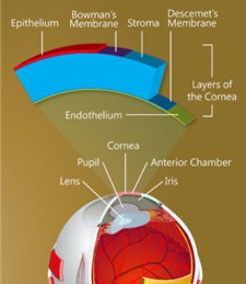
Illustration of structure of cornea

The structure of the cornea.
(Keratomania.com eye diagram by
Chabacano,via Wikimedia Commons.)

On the backside of the cornea is a single layer of cells that plays an all-important role, maintaining just the right fluid balance to keep the cornea transparent so that light can enter the eye. Until recently, it was believed this layer, called the corneal endothelium, is incapable of replacing its damaged cells. As more cells become damaged, the cornea becomes opaque, leading to loss of vision and, ultimately, to as many as 30,000 endothelium transplants a year in the United States alone.

A team of University researchers is exploring the possibility that stem cells on the outer edges of the cornea, given the right stimulation, can migrate into the endothelium to replace damaged cells. (Undifferentiated stem cells develop into specialized cells.) The work raises the possibility of restoring vision without the need for transplants.

The team is led by Amy Kiernan, associate professor of ophthalmology, and includes Jannick Rolland, the Brian J. Thompson Professor of Optical Engineering; Patrice Tankam, a senior scientist in the Center for Visual Science; Changsik Yoon, a graduate student in Rolland's lab; Rebecca Rausch, a graduate student in Kiernan's lab; and Holly Hindman, former associate professor of ophthalmology, now in private practice but still consulting on the project. They are supported with a $75,000 University Research Award. The URA program is designed to help researchers develop preliminary data or proof of concept needed to leverage larger federal or foundation awards to carry a promising project to completion.

There have been tantalizing clinical hints that the corneal endothelium may have regenerative capabilities, Kiernan says. For example, there have been cases in which endothelial transplants failed to engraft, but the cornea cleared up anyway, with regeneration of the endothelium occurring on its own. "So it seems that if something is done that stimulates a progenitor or stem cell population, most likely those in the periphery of the cornea, there is some regenerative capacity in the endothelium -- just based on clinical studies," Kiernan says.

Her team will attempt to identify the potential stem cells that might be stimulated to migrate to the endothelium to repair damage. They will use mouse models from Kiernan's lab in which adult stem cells can be permanently tagged with fluorescent biomarkers and tracked even after they differentiate into other cells. The identification and tracking of those cells will be done by refining a novel imaging approach developed in Rolland's lab. Called Gabor domain optical coherence microscopy, the technology allows rapid, noninvasive imaging of cellular structures beneath the surface of the skin or within the human eye -- in greater detail than traditional imaging with optical coherence tomography.

"Think of it as a high-definition, volumetric imaging," Rolland says. "But we also want to know what kind of cells we are looking at, so we are integrating fluorescence imaging with the high-definition volumetric microscopy so we can do both." The team represents a combination of pertinent expertise: cell development and regeneration (Kiernan and Rausch), imaging (Rolland, Tankam, and Yoon), and the biological basis for corneal and ocular surface diseases in humans (Hindman). The University Research Award funding is helping support graduate students and technicians working on the project, and the cost of mice and supplies. "Pilot funding like this is so important, especially with NIH grants shrinking," Kiernan says.

"It's really helpful to be able to bridge this kind of interdisciplinary effort," says Rolland. "You need to work together a little bit to understand the challenges involved and what you need to do to secure preliminary data, to show we have a pathway. "It takes time to get data, so even a small grant that provides a bridge for a year or two can make a huge difference."

Nina Schor to Step Down as Children's Hospital Pediatrician-in-Chief

Wednesday, December 7, 2016

Nina Schor, M.D., Ph.D., William H. Eilinger Chair of Pediatrics at the University of Rochester Medical Center (URMC), will step down as chair in June 2017. Schor served 11 years as pediatrician-in-chief at UR Medicine’s Golisano Children’s Hospital, and under her leadership, the Department of Pediatrics fulfilled a decades-long dream of building a standalone children’s hospital in Rochester; the new facility opened its doors to patients in July 2015. "I don’t want to downplay the significance of the new hospital, but it’s really what we do inside of it and because of it that’s so important,” said Schor. “I look at the academic physicians and physician scientists who came to Rochester with just a dream and a fire in their belly and how they’ve now brought those dreams to fruition — that’s what I’m most proud of.”

The Department of Pediatrics grew from 110 faculty members to over 170 during Schor’s tenure, creating new divisions in palliative care, sleep medicine, allergy, and hospitalist medicine. Research centers focused on premature infants, translational molecular programs, and red blood cell development also developed under Schor’s leadership.

“Not only was the new hospital built under Nina’s leadership, but she truly championed the project, ensuring that every detail was designed with patients and families in mind,” said Mark Taubman, M.D., URMC CEO and Dean of the School of Medicine and Dentistry. “She has been the face of the children’s hospital and inspired trust in our families, physicians, and donors at a time when we very much needed the community’s support.”

Read More: Nina Schor to Step Down as Children's Hospital Pediatrician-in-Chief

Study Challenges Autism Brain Response Theory

Tuesday, December 6, 2016

A new study challenges the hypothesis that nerve cells in the brains of individuals with Autism Spectrum Disorders do not reliably and consistently respond to external stimuli. “Our findings show there is no measurable variation in how individuals with autism respond to repeated visual and tactile stimuli,” says senior author John Foxe, the Kilian J. and Caroline F. Schmitt Professor in Neuroscience.

Read More: Study Challenges Autism Brain Response Theory

NIH Pre-Doctoral Fellowship Award

Tuesday, November 29, 2016

Matt Cavanaugh, a fifth year Neuroscience Graduate Program student in Dr. Krystel Huxlin’s lab was awarded an NIH Individual Pre-doctoral Fellowship from the National Eye Institute for his project entitled: Properties of training-induced visual recovery in cortical blindness (2016-2019).

Congratulations Matt!

Brain training video games help low-vision kids see better

Monday, November 28, 2016

Studies going back several years have shown that playing action video games (AVG) can help improve visual acuity. A new study by vision scientists at the University of Rochester and Vanderbilt University found that children with poor vision see vast improvement in their peripheral vision after only eight hours of training via kid-friendly video games. Most surprising to the scientists was the range of visual gains the children made, and that the gains were quickly acquired and stable when tested a year later.

"Children who have profound visual deficits often expend a disproportionate amount of effort trying to see straight ahead, and as a consequence they neglect their peripheral vision," said Duje Tadin, associate professor of brain and cognitive sciences at Rochester. "This is problematic because visual periphery—which plays a critical role in mobility and other key visual functions—is often less affected by visual impairments."

"We know that action video games (AVG) can improve visual perception, so we isolated the AVG components that we thought would have the strongest effect on perception and devised a kid-friendly game that compels players to pay attention to the entire visual field, not just where their vision is most impaired," said Tadin, who is also a professor in the Center for Visual Science. "As a result, we've seen up to 50 percent improvement in visual perception tasks."

Read More: Brain training video games help low-vision kids see better

Children’s Hospital Pediatrician-in-Chief Named Fellow of the AAAS

Monday, November 21, 2016

Nina Schor, M.D., Ph.D., William H. Eilinger Chair of Pediatrics and the pediatrician-in-chief at UR Medicine’s Golisano Children’s Hospital, has been named a Fellow of the American Association for the Advancement of Science (AAAS), the world’s largest general scientific society.

AAAS, which will publish the announcement on Nov. 25 in its journal Science, selects Fellows based on their scientifically and socially distinguished efforts to advance science or its applications. Schor has spent much of her career investigating neuroblastoma — which is among the most common childhood cancers — and was recognized for “her distinguished contributions to developmental neuroscience and neuropharmacology, particularly using molecular neuroscientific discoveries to design innovative therapies for tumors of the developing nervous system.”

Read More: Children’s Hospital Pediatrician-in-Chief Named Fellow of the AAAS

Repurposed Drug May Offer Diagnosis, Treatment for Traumatic Nerve Damage

Monday, November 14, 2016

Researchers at the University of Rochester Medical Center believe they have identified a new means of enhancing the body's ability to repair its own cells, which they hope will lead to better diagnosis and treatment of traumatic nerve injuries, like those sustained in car accidents, sports injuries, or in combat. In a study published today, the team showed that a drug previously approved for other purposes can 'wake up' damaged peripheral nerves and speed repair and functional recovery after injury.

The study appearing in EMBO Molecular Medicine, demonstrates for the first time that 4-aminopyridine (4AP), a drug currently used to treat patients with the chronic nerve disease, multiple sclerosis, has the unexpected property of promoting recovery from acute nerve damage. Although this drug has been studied for over 30 years for its ability to treat chronic diseases, this is the first demonstration of 4AP's benefit in treating acute nerve injury and the first time those benefits were shown to persist after treatment was stopped.

Study authors, Dr. John Elfar, associate professor of Orthopaedics, and Mark Noble, Ph.D., Martha M. Freeman, M.D., Professor in Biomedical Genetics, and their laboratory team, found that daily treatment with 4AP promotes repair of myelin, the insulating material that normally surrounds nerve fibers, in mice. When this insulation is damaged, as occurs in traumatic peripheral nerve injury, nerve cell function is impaired. These researchers found that 4AP treatment accelerates repair of myelin damage and improvement in nerve function.

Read More: Repurposed Drug May Offer Diagnosis, Treatment for Traumatic Nerve Damage

Research Will Explore New Therapies for Huntington’s Disease

Wednesday, October 12, 2016

Image of astocytes

A new award from the CHDI Foundation will advance promising research that aims to slow the progression of Huntington's disease. The funding, anticipated to total more than $10.5 million over next five years, will help University of Rochester Medical Center (URMC) scientists develop a stem cell-based therapy that swaps sick brain cells for healthy ones.

The new award will go to the lab of Steve Goldman, M.D., Ph.D., the co-director of the URMC Center for Translational Neuromedicine, which has research operations in both Rochester and at the University of Copenhagen.

Huntington's is a hereditary neurodegenerative disease characterized by the loss of medium spiny neurons, a nerve cell in the brain that plays a critical role in motor control. As the disease progresses over time and more of these cells die, the result is involuntary movements, problems with coordination, and cognitive decline, depression, and often psychosis. There is currently no way to slow or modify this fatal disease.

The new award will support research that builds upon findings published by Goldman earlier this year in the journal Nature Communications showing that researchers were able to slow the progression of the disease in mice by transplanting healthy human support cells, called glial progenitor cells, into the animals' brains.

Read More: Research Will Explore New Therapies for Huntington’s Disease

Armond Collins Presents his work from Fudge Lab

Tuesday, October 11, 2016

Photo of Armond Collins

Armond Collins, a second year medical student, presents the study he conducted with us this summer under the auspices of a Babigian Fellowship. Armond studied changes in myelination in amygdala and cortex of adult rats that had been exposed to 3 day bout of a repeated variable stressor during adolescence. His works follows up studies by Michele Saul, PhD that indicate that adolescent stress results in decreased oligodendrocyte precursors in the amygdala in the week following the stress.

New Grants Explore Role of Brain’s “Garbage Truck” in Mini-Stokes and Trauma

Wednesday, October 5, 2016

Photo of Dr. Nedergaard

Maiken Nedergaard, M.D., D.M.Sc.

More than $4.5 million in new grants to the lab of University of Rochester Medical Center scientist Maiken Nedergaard, M.D., D.M.Sc., underscore the important role the brain's waste disposal system may play in a range of neurological disorders. The new awards will advance understanding of how small vessel disease and traumatic brain injury can give rise to cognitive and behavioral problems.

Nedergaard and her colleagues first unveiled the brain's unique method of removing waste -- dubbed the glymphatic system -- in a paper in Science Translational Medicine in 2012. The research revealed that the brain possesses a circulation network that piggybacks on blood vessels and uses cerebral spinal fluid to flush away waste products from brain tissue. Since that time, the team has gone on to show that the glymphatic system works primarily while we sleep, could be a key player in diseases like Alzheimer's, and is disrupted after traumatic brain injury.

Read More: New Grants Explore Role of Brain’s “Garbage Truck” in Mini-Stokes and Trauma

NGP Student Awarded NIH Fellowship

Monday, October 3, 2016

Rebecca Rausch, a fifth year neuroscience graduate student in Dr. Richard Libby's lab was awarded an NIH Individual Pre-doctoral Fellowship from the National Eye Institute for her project entitled: The Role of Notch and BMP Signaling in Anterior Segment Dysgenesis (2016-2019).

Congratulations Becca!

Nuclear Protein Causes Neuroblastoma to Become More Aggressive

Wednesday, September 28, 2016

Image of Photoblastomas

Aggressive forms of neuroblastoma contain a specific protein in their cells’ nuclei that is not found in the nuclei of more benign forms of the cancer, and the discovery, made through research from the University of Rochester Medical Center (URMC), could lead to new forms of targeted therapy.

EYA1, a protein that contributes to ear development, is present in the cytoplasm of many neuroblastoma tumors, but this protein migrates to the nucleus in the cells of more aggressive forms of the disease. The research, recently published in two medical research journals, allows for the development of targeted drugs that will work to prevent the neuroblastoma from reaching this more aggressive stage; researchers at URMC and elsewhere have already begun testing some of these potential treatments in a laboratory setting.

Photo of Nina Schor

“Neuroblastoma is one of the most common and deadly forms of childhood cancer, and this discovery allows us to identify drugs that prevent the change in EYA1 structure and potentially minimize the danger to a child who has this disease,” said Nina Schor, M.D., Ph.D., professor of Pediatrics and Neuroscience and the William H. Eilinger Chair of Pediatrics at URMC.

Read More: Nuclear Protein Causes Neuroblastoma to Become More Aggressive

Jessica Cantlon Named One of 10 Scientists to Watch by Science News

Friday, September 23, 2016

Photo of Jessica Cantlon

Jessica Cantlon

Jessica Cantlon, associate professor of Brain and Cognitive Sciences, was selected by Science News as one of their 10 early- to mid-career scientists to watch. Cantlon’s work centers on how human and nonhuman primates distinguish between quantities. Understanding how the brain makes sense of concepts such as estimating quantities and counting might lead to better ways of teaching numerical concepts to children.

Read More: Jessica Cantlon Named One of 10 Scientists to Watch by Science News

Harris Gelbard Receives International Award for Neurovirology Research

Tuesday, September 13, 2016

Harris GelbardHarris "Handy" Gelbard, M.D., Ph.D., director of the Center for Neural Development & Disease, is slated to receive the Hilary Koprowski Prize in Neurovirology at this year's International Symposium on Molecular Medicine and Infectious Disease at Drexel University. Gelbard will be recognized for developing an unconventional drug that shows promise in treating brain disorders associated with HIV.

Gelbard's drug, URMC-099, calms the immune system when it goes awry, as happens in HIV Associated Neurocognitive Disorder (HAND). In HAND, immune reactions to HIV particles in the brain damage nerve cells and cause dementia. Because patients affected by HAND also have HIV, it was imperative that URMC-099 not interfere with the antiretroviral drugs that keep HIV-positive patients alive.

2016 Convocation Award Winners from Neuroscience

Thursday, September 8, 2016

Congratulations to the following people for winning teaching and student achievement awards at this year's SMD Opening Convocation.

Faculty Teaching, Mentoring & Diversity Awards

  • Deborah Cory-Slechta, PhD
  • John Olschowka, PhD

Medical & Graduate Student Achievement Awards

  • Alexandra McHale - Irving L. Spar Fellowship Award
  • Gavin Jenkins - Merritt and Marjorie Cleveland Fellowship
  • Neal Shah - J. Newell Stannard Graduate Student Scholarship Award
  • Grayson Sipe - Outstanding Student Mentor Award

Make sure to congratulate each of them when you see them.

DOD Grant Explores New Drugs to Thwart Impact of Trauma, Stroke, and Cardiac Arrest

Thursday, September 8, 2016

Photo of soldier in desert

A $2.3 million Department of Defense grant will help neuroscientists develop new treatments for the emergency room and the battlefield. The research will focus on the development of new therapies that could help protect brain and other at risk organs following a trauma, heart attack, or stroke.

“While we have made significant progress in our ability to restore blood flow after stroke or cardiac arrest, the medical community does not have drugs at its disposal to prevent the secondary damage that occurs after these events,” said University of Rochester Medical Center neurologist Marc Halterman, M.D., Ph.D., the principal investigator of the study. “This grant will further our research on a promising class of drugs that possess both anti-inflammatory and cytoprotective properties that we believe will be suitable for use in both military and emergency conditions.”

Read More: DOD Grant Explores New Drugs to Thwart Impact of Trauma, Stroke, and Cardiac Arrest

How Does Noise Damage Hearing?

Tuesday, September 6, 2016

Join Pat White on Saturday, September 10 from 2-4pm at the Pittsford Barnes and Noble as she is featured at the UR Science Cafe discussion

Automatic cortical representation of auditory pitch changes in Rett syndrome - John Foxe et al.

Thursday, September 1, 2016

Over the typical course of Rett syndrome, initial language and communication abilities deteriorate dramatically between the ages of 1 and 4 years, and a majority of these children go on to lose all oral communication abilities. It becomes extremely difficult for clinicians and caretakers to accurately assess the level of preserved auditory functioning in these children, an issue of obvious clinical import. Non-invasive electrophysiological techniques allow for the interrogation of auditory cortical processing without the need for overt behavioral responses. In particular, the mismatch negativity (MMN) component of the auditory evoked potential (AEP) provides an excellent and robust dependent measure of change detection and auditory sensory memory. Here, we asked whether females with Rett syndrome would produce the MMN to occasional changes in pitch in a regularly occurring stream of auditory tones.

Read More: Automatic cortical representation of auditory pitch changes in Rett syndrome - John Foxe et al.

Lin Honored as 'Brilliant New Investigator'

Thursday, August 4, 2016

Photo of Dr. LinUniversity of Rochester Assistant Professor of Nursing Feng (Vankee) Lin, Ph.D., R.N. will be presented with the Brilliant New Investigator Award from the Council for the Advancement of Nursing Science (CANS) at the organization’s 2016 State of the Science Congress on Nursing Research, Sept. 15-17 in Washington, D.C.

The award recognizes the contributions of scientists early in their research careers who show extraordinary potential to develop sustained programs of research certain to have significant impact on the science and practice of nursing and health care. Nominees must show a record of building research productivity in an area of major significance to nursing and health care, research dissemination and translation to practice and/or policy, and emerging leadership related to the advancement of nursing science.

Read More: Lin Honored as 'Brilliant New Investigator'

Pasternak Research Paper to be Published in J. Neuroscience

Thursday, August 4, 2016

Photo of Tania PasternakThe paper "Prefrontal Neurons Represent Motion Signals from Across the Visual Field but for Memory-Guided Comparisons Depend on Neurons Providing these Signals" will be published in J. Neuroscience shortly.

Visual decisions often involve comparisons of sequential visual motion that can appear at any location in the visual field. We show that during such comparisons, the lateral prefrontal cortex (LPFC) contains accurate representation of visual motion from across the visual field, supplied by motion processing neurons. However, at the time of comparison, LPFC neurons can only use this information to compute the differences between the stimuli, if stimuli appear at the same retinal location, implicating neurons with localized receptive fields in the comparison process. These findings show that sensory comparisons rely on the interactions between LPFC and sensory neurons that not only supply sensory signals but also actively participate in the comparison of these signals at the time of the decision.

Make sure to read the article when it comes out.

Luebke and Bennetto Explore Hearing Test That May Identify Autism Risk

Monday, July 25, 2016

Diagram of hearing test to identify autism risk

Researchers have identified an inner ear deficiency in children with Autism that may impact their ability to recognize speech. The findings, which were published in the journal Autism Research, could ultimately be used as a way to identify children at risk for the disorder at an early age.

“This study identifies a simple, safe, and non-invasive method to screen young children for hearing deficits that are associated with Autism,” said Anne Luebke, Ph.D., an associate professor in the University of Rochester Medical Center Departments of Biomedical Engineering and Neuroscience and a co-author of the study. “This technique may provide clinicians a new window into the disorder and enable us to intervene earlier and help achieve optimal outcomes.”

“Auditory impairment has long been associated with developmental delay and other problems, such as language deficits,” said Loisa Bennetto, Ph.D., an associate professor in the University of Rochester Department of Clinical and Social Sciences in Psychology and a co-author of the study. “While there is no association between hearing problems and autism, difficulty in processing speech may contribute to some of the core symptoms of the disease. Early detection could help identify risk for ASD and enable clinicians to intervene earlier. Additionally, these findings can inform the development of approaches to correct auditory impairment with hearing aids or other devices that can improve the range of sounds the ear can process.”

Read More: Luebke and Bennetto Explore Hearing Test That May Identify Autism Risk

McNair Summer Scholar Ashley Bui Talks Amygdala Circuits

Friday, July 22, 2016

Photo of Ashley BuiCongratulations to Ashley Bui, a rising senior in Brain and Cognitive Sciences, on her presentation July 22, 2016. Her talk Projections from the Temporal Cortex to the Basal Nucleus of the Amygdala in the Macaque highlighted data from her summer project in our lab. The amygdala is required for computing which of the complex sensory stimuli that an individual encounters are emotionally meaningful, so that appropriate action can be taken. Ashley’s preliminary data shows that specific portions of the temporal cortex, which are critical for processing complex visual and auditory information, communicate with different regions of the amygdala. The results suggest that cortical areas that process complex visual information on 'what' and 'where' an object is (or is moving) are communicating with specific amygdala subregions. Thus, while determining the emotional importance of ‘what or who’ is important, biologic movements also likely influence amygdala activity and coding. We are happy that she will continue this work through the Fall semester.

NGP student plays with RPO

Thursday, July 21, 2016

Monique Mendes plays with RPO

Second year NGP student, Monique Mendes, had a unique opportunity to play alongside the Rochester Philharmonic Orchestra in their Side-by-Side Reading Session – a program that pairs amateur and professional musicians in a joint rehearsal and performance at Kodak Hall on July 21st.
Congratulations Monique!

Ryan Dawes defends thesis

Monday, July 18, 2016

Photo of Ryan Dawes

Ryan Dawes successfully defended his thesis, "β-Adrenergic Receptor Signaling Constrains Breast Cancer Progression and Modulates Tumor-Associated Exosome Content And Function" on July 18, 2016.

Congratulations Dr. Dawes!

Rebecca Lowery Defends Thesis

Thursday, July 7, 2016

Rebecca Lowery and Ania Majewska

Rebecca Lowery has successfully defended her thesis, "The Role of Microglia and Fractalkine Signaling in Experience-dependent Synaptic Plasticity". Congratulate her when you see her.

Congratulations Dr. Lowery!

The Sleep Hack Neuroscience Says Gives Your Brain Optimal Rest

Thursday, June 30, 2016

Sleep is critical for rest and rejuvenation. A human being will actually die of sleep deprivation before starvation--it takes about two weeks to starve, but only 10 days to die if you go without sleep.

The CDC has also classified insufficient sleep as a public health concern. Those who don't get enough sleep are more likely to suffer from chronic diseases that include hypertension, diabetes, depression, obesity, and cancer.

It's thus vital to get enough shuteye, but it turns out your sleep position also has a significant impact on the quality of rest you get.

Now, a neuroscience study suggests that of all sleep positions, one is most helpful when it comes to efficiently cleaning out waste from the brain: sleeping on your side.

The study, published in the Journal of Neuroscience, used dynamic contrast-enhanced MRI to image the brain's "glymphatic pathway." This is the system by which cerebrospinal fluid filters through the brain and swaps with interstitial fluid (the fluid around all other cells in the body).

"It is interesting that the lateral [side] sleep position is already the most popular in humans and most animals--even in the wild," said University of Rochester's Maiken Nedergaard. "It appears that we have adapted the lateral sleep position to most efficiently clear our brain of the metabolic waste products that build up while we are awake."

Read More: The Sleep Hack Neuroscience Says Gives Your Brain Optimal Rest

Elissa Wong receives Neuman Scholarship Award

Tuesday, June 21, 2016

Elissa Wong being presented with the Newman Award

Elissa Wong, a fifth year toxicology graduate student in Ania Majewska’s lab, received the Margaret and William F. Neuman Scholarship Award in Environmental Medicine for exemplary scholarship and citizenship. Dr. William Neuman was the chair of the Department of Radiation Biology and Biophysics for many years and helped to create the Toxicology Training Program and the Environmental Health Science Center. Dr. Margaret Neuman received her PhD in Biochemistry from the University of Rochester. Later, working here, she researched the effects of uranium on bone biochemistry, and was an expert on the regulation of bone minerals.

The criteria for receiving this are as follows: 1) scholarship, 2) scientific excellence, 3) productivity, and 4) exceptional citizenship to the field of toxicology.

Congratulations Elissa!

Swapping Sick for Healthy Brain Cells Slows Huntington’s Disease

Tuesday, June 7, 2016

Photo of Steven Goldman

Researchers have successfully reduced the symptoms and slowed the progression of Huntington’s disease in mice using healthy human brain cells. The findings, which were published today in the journal Nature Communications, could ultimately point to a new method to treat the disease.

The research entailed implanting the animals with human glia cells derived from stem cells. One of the roles of glia, an important support cell found in the brain, is to tend to the health of neurons and the study’s findings show that replacing sick mouse glia with healthy human cells blunted the progress of the disease and rescued nerve cells at risk of death.

“The role that glia cells play in the progression of Huntington’s disease has never really been explored,” said Steve Goldman, M.D., Ph.D., co-director of the University of Rochester Center for Translational Neuromedicine. “This study shows that these cells are not only important actors in the disease, but may also hold the key to new treatment strategies.”

Read More: Swapping Sick for Healthy Brain Cells Slows Huntington’s Disease

Proposal by Amy Kiernan Receives University Research Award

Monday, May 23, 2016

A collaborative project involving Associate Professor Amy Kiernan of the Flaum Eye Institute has been chosen as one of the 2016-17 University Research Awards. One of just eight applications chosen by senior research leadership, the proposal entitled, "Understanding cell turnover and injury recovery in the corneal endothelium" will be funded $75.000 annually.

Conventional Radiation Therapy May Not Protect Healthy Brain Cells

Wednesday, May 18, 2016

Photo demo of conventional radiation therapy

A new study shows that repeated radiation therapy used to target tumors in the brain may not be as safe to healthy brain cells as previously assumed. The findings, which appear in the International Journal of Radiation Oncology, Biology, Physics, show that the treatment also kills important support cells in the brain and may cause as much, if not more damage, than single dose radiation therapy.

“This study suggests that conventional repeated radiation treatments offer no significant benefit to brain tumor patients,” said Kerry O’Banion, M.D., Ph.D., a professor in the University of Rochester Medical Center (URMC) Department of Neuroscience and lead author of the study. “It also shows that certain cell populations in the brain are vulnerable to radiation and this may help explain why so many brain cancer patients experience cognitive problems after treatment.”

Read More: Conventional Radiation Therapy May Not Protect Healthy Brain Cells

When the Physical World is Unreliable: Study Finds Visual and Tactile Processing Deficits in Schizophrenia

Tuesday, May 10, 2016

Schizophrenia

A new study out today in the journal Translational Psychiatry sheds further light on the idea that schizophrenia is a sensory disorder and that individuals with the condition are impaired in their ability to process stimuli from the outside world. The findings may also point to a new way to identify the disease at an early stage and before symptoms become acute.

Because one of the hallmarks of the disease is auditory hallucinations, such as hearing voices, researchers have long suspected a link between auditory processing and schizophrenia. The new study provides evidence that the filtering of incoming visual information, and also of simple touch inputs, is also severely compromised in individuals with the condition.

“When we think about schizophrenia, the first things that come to mind are the paranoia, the delusions, the disorganized thinking,” said John Foxe, Ph.D., the chair of the University of Rochester Medical Center Department of Neuroscience and senior author of the study. “But there is increasing evidence that there is something fundamentally wrong with the way these patients hear, the way they feel things through their sense of touch, and in the way in which they see the environment.”

Read More: When the Physical World is Unreliable: Study Finds Visual and Tactile Processing Deficits in Schizophrenia

SA Government names Professors of the Year

Monday, May 2, 2016

Laurel Carney

Join us in congratulating Laurel for being selected as one of 4 Professors of the year from a extraordinary field of 63 candidates.

Laurel Carney, professor of biomedical engineering, won in the Engineering field. Her research focuses on the complex network of auditory nerve fibers that transmit the inner ear’s electrical signals to the brain with the goal of better hearing aids.

Carney earned her M.S. and Ph.D degrees in electrical engineering at the University of Wisconsin-Madison. She was an associate professor of biomedical engineering at Boston University and professor of biomedical engineering at Syracuse University before joining the Rochester faulty in 2007. She serves as professor in three departments – biomedical engineering, neurobiology and anatomy, and electrical and computer engineering.

Read More: SA Government names Professors of the Year

Elissa Wong Awarded Individual Pre-doctoral Fellowship from NIAAA

Sunday, May 1, 2016

Photo of Elissa Wong

Elissa Wong, a 4th year Toxicology Graduate Program student in Dr. Ania Majewska's lab received a perfect 10 review score and was awarded an NIH (NRSA) Individual Pre-doctoral Fellowship from the NIAAA. The title of her project is: Synaptic plasticity and microglial-synapse interactions after developmental alcohol exposure (2016-2018).
Congrats Elissa!

NGP Graduate Alum, Grayson Sipe, Wins Doty Thesis Award

Friday, April 29, 2016

Photo of Grayson Sipe

Grayson Sipe, recent doctoral graduate from the Majewska lab, received the Robert Doty prize for the 2016 outstanding dissertation in neuroscience. The Doty prize is named in the honor of longtime faculty member Robert Doty, who made great contributions to neuroscience research at the University of Rochester and nationally. It is awarded on the basis of the impact and importance of research, novelty of experimental design, independence and creativity of the student and research implications and relevance for neuroscience. Grayson’s thesis entitled “The Role of P2Y12 in non-pathological microglial functions during synaptic plasticity”, which he successfully defended on February 19th, 2016, embodied all these characteristics. Grayson has now moved to his postdoctoral position with Dr. Mriganka Sur at MIT. Dr. Peter Shrager presented Grayson the prize at the annual neuroscience retreat on Friday, April 29th.

Congratulations Grayson!!!

Subtle Chemical Changes in Brain Can Alter Sleep-Wake Cycle

Friday, April 29, 2016

Sleepy Brain

A study out today in the journal Science sheds new light on the biological mechanisms that control the sleep-wake cycle. Specifically, it shows that a simple shift in the balance of chemicals found in the fluid that bathes and surrounds brain cells can alter the state of consciousness of animals.

The study, which focuses on a collection of ions that reside in the cerebral spinal fluid (CSF), found that not only do these changes play a key role in stimulating or dampening the activity of nerve cells, but they also appear to alter cell volume causing brain cells to shrink while we sleep, a process that facilitates the removal of waste.

"Understanding what drives arousal is essential to deciphering consciousness and the lack thereof during sleep," said Maiken Nedergaard, M.D., D.M.Sc., co-director of the University of Rochester Center for Translational Neuromedicine and lead author of the study. "We found that the transition from wakefulness to sleep is accompanied by a marked and sustained change in the concentration of key extracellular ions and the volume of the extracellular space."

The current scientific consensus is that the brain is "woken up" by a set of neurotransmitters -- which include compounds such as acetylcholine, hypocretin, histamine, serotonin, noradrenaline, and dopamine -- that originate from structures deep within the brain and the brain stem. This cocktail of chemical messengers serve to activate -- or arouse -- a set of neurons in the cerebral cortex and other parts of the brain responsible for memory, thinking, and learning, placing the brain in a state of wakefulness.

Read More: Subtle Chemical Changes in Brain Can Alter Sleep-Wake Cycle

Congratulations to NGP student Aleta Steevens

Friday, April 8, 2016

Photo of Aleta Steevens

Aleta Stevens, an NGP student in Dr. Amy Kiernan's lab, secured a 3-year NIH Individual Pre-doctoral Fellowship, F31 entitled, "Elucidating the role of SOX2 in inner ear development."

Excellent work Aleta!

Neuroscience Graduate Students Win Award for Teaching

Friday, April 8, 2016

Aleta Steevens

Neuroscience Graduate Program students, Aleta Steevens (Dr. Amy Kiernan lab) and Heather Natola (Dr. Chris Pröschel lab) were awarded the 2016 Edward Peck Curtis Award for Excellence for Graduate Student Teaching.

Only a handful of these are awarded each year, and all this year's nominees were extremely well-qualified.

Congratulations to both!!!

Christina Cloninger Defends Thesis

Thursday, April 7, 2016

Photo of Christina CloningerCongratulations Dr. Cloninger on successfully defending your thesis!!

“Honeycomb” of Nanotubes Could Boost Genetic Engineering

Wednesday, April 6, 2016

Electron microscopic image of animal cells on array of nanotubes

Electron microscope image of animal cells (colored blue) cultured on an array of carbon nanotubes

Researchers have developed a new and highly efficient method for gene transfer. The technique, which involves culturing and transfecting cells with genetic material on an array of carbon nanotubes, appears to overcome the limitations of other gene editing technologies.

The device, which is described in a study published today in the journal Small, is the product of a collaboration between researchers at the University of Rochester Medical Center (URMC) and the Rochester Institute of Technology (RIT).

“This platform holds the potential to make the gene transfer process more robust and decrease toxic effects, while increasing amount and diversity of genetic cargo we can deliver into cells,” said Ian Dickerson, Ph.D., an associate professor in the Department of Neuroscience at the URMC and co-author of the paper.

Read More: “Honeycomb” of Nanotubes Could Boost Genetic Engineering

Early Wiring of Brain's “Fear” Centers Could Produce Long-term Consequences

Monday, March 21, 2016

Fear from Early Brain Wiring

New research shows that our brains may be hardwired to become sensitive to stressful environments at an early age and, if overstimulated, this may contribute to anxiety disorders and even psychotic syndromes later in life.

The study, which appears in the journal Brain Structure and Function, focuses on two structures deep in the brain. The central nucleus of the amygdala (Ce) is thought to be involved in responses to immediate threats and stimulus, such as becoming startled or freezing in reaction to a loud noise. The bed nucleus of the stria terminalis (BST) is thought to be involved in regulating a person’s state of vigilance, such as determining whether or not an environment or a situation poses a potential threat. Animal and human studies show that when the BST is activated by a threatening situation, we tend to slow down, become quieter, and stress hormones spike.

While Ce and BST reside in different parts of the brain, the two areas are hardwired to each other by axonal tracts – basically, bundles of long distance axon fibers that enable the separate regions to communicate with each other. However, until now it has not been clear when these connections form or the way in which they interact with each other.

In the study published today, a team of researchers led by Julie Fudge, M.D., with the Department of Neuroscience observed that these connections are made at a very early stage of development in non-human primates. They also found that the direction of the connection is essentially a one way street. The Ce – or immediate fear signaling center – conveys information to the BST, the structure that mediates general threat sensing or anxiety states. This arrangement suggests that repeated activation of the Ce by immediately fearful or traumatic events may shape long-term anxiety states in the BST.

Read More: Early Wiring of Brain's “Fear” Centers Could Produce Long-term Consequences

The Brain’s Gardeners: Immune Cells ‘Prune’ Connections Between Neurons

Monday, March 7, 2016

MicrogliaMicroglia (green) with purple representing the P2Y12 receptor which the study shows is a critical regulator in the process of pruning connections between nerve cells.

A new study out today in the journal Nature Communications shows that cells normally associated with protecting the brain from infection and injury also play an important role in rewiring the connections between nerve cells. While this discovery sheds new light on the mechanics of neuroplasticity, it could also help explain diseases like autism spectrum disorders, schizophrenia, and dementia, which may arise when this process breaks down and connections between brain cells are not formed or removed correctly.

“We have long considered the reorganization of the brain’s network of connections as solely the domain of neurons,” said Ania Majewska, Ph.D., an associate professor in the Department of Neuroscience at the University of Rochester Medical Center (URMC) and senior author of the study. “These findings show that a precisely choreographed interaction between multiple cells types is necessary to carry out the formation and destruction of connections that allow proper signaling in the brain.”

The study is another example of a dramatic shift in scientists’ understanding of the role that the immune system, specifically cells called microglia, plays in maintaining brain function. Microglia have been long understood to be the sentinels of the central nervous system, patrolling the brain and spinal cord and springing into action to stamp out infections or gobble up dead cell tissue. However, scientists are now beginning to appreciate that, in addition to serving as the brain’s first line of defense, these cells also have a nurturing side, particularly as it relates to the connections between neurons.

Read More: The Brain’s Gardeners: Immune Cells ‘Prune’ Connections Between Neurons

Hope, Hype, and Wishful Thinking

Monday, February 22, 2016

Dr. Goldman

In a perspective piece appearing in the journal Cell Stem Cell, URMC neurologist Steve Goldman, M.D., Ph.D., lays out the current state of affairs with respect to stem cell medicine and how close we are to new therapies for neurological disorders.

The dawn of stem cell medicine some 25 years ago was greeted with great enthusiasm, particularly by scientists who study diseases in the central nervous system (CNS). Many of the diseases found in the brain and spinal cord are degenerative in nature; meaning that over time populations of cells are lost due to genetic factors, infection, or injury. Because stem cell medicine holds the potential to repair or replace damaged or destroyed cells, scientists have considered these diseases as promising candidates for new therapies.

However, as with other emerging fields of medicine, the race to cures has turned out to be more of marathon than a sprint. While scientists have become very adept at manipulating stem and progenitor cells and understanding the complex choreography of genetic and chemical signals that instruct these cells to divide, differentiate, and proliferate, researchers are still grappling with the challenges of how to integrate new cells into the complex network of connections that comprise the human brain.

Goldman, co-director of the URMC Center for Translational Neuromedicine, takes a sweeping view of where we stand and which CNS diseases may or may not ultimately benefit from future stem cell-based therapies.

Read More: Hope, Hype, and Wishful Thinking

Congratulations Dr. Sipe!

Wednesday, February 17, 2016

Make sure you congratulate Grayson Sipe on defending his thesis.
Way to go Grayson!

Richard Aslin's Rochester Baby Lab Shows

Wednesday, February 17, 2016

Amelia Smith sits on the floor of a newly remodeled wing of the University of Rochester's department of Brain and Cognitive Sciences. The 8-month old wears a headband of cottony roses, and tiny bubbles form in the corner of her mouth. She's completely entranced by the commotion around her.

Though few adults in the room can resist oohing and aww-ing, little Amelia is not there to be fawned over. She's there to work. Researchers at the UR's Baby Lab want to know what she's thinking, what she's learned so far in her young life, and how she learned it.

But there's a problem: Amelia can't talk yet.

The work being investigated in Richard Aslin's Baby Lab was written up in the City Newspaper article "Signs of Intelligent Life".

Read More: Richard Aslin's Rochester Baby Lab Shows

Report Recommends More Treatment, Research, for Gulf War Vets

Monday, February 15, 2016

The cause of Gulf War illness is still a mystery but focusing on treatments and interventions might help the veterans of Operation Desert Storm as well as the troops of the future, according to an Institute of Medicine committee report led by University of Rochester Medical Center Professor Deborah Cory-Slechta.

In 1990 and ’91 nearly 700,000 U.S. troops deployed to the Persian Gulf region for a short, intense war. Few injuries or deaths occurred, but troops were exposed to chemical and biological weapons, vaccines, oil-fires, air pollution, bomb blasts, pesticides, extreme desert temperatures, and constant false alarms and fear of nerve-gas attacks.

After the war ended a high number of the veterans reported debilitating fatigue, muscle and joint pain, headaches, and cognitive problems. This became known as “Gulf War illness.” During the past 25 years, 10 different committees of the nation’s top medical experts have searched for evidence that would better define Gulf War illness and possible treatments. The latest committee, headed by Cory-Slechta, concluded that no single mechanism can explain the multitude of symptoms seen in Gulf War illness—and that it’s unlikely a cause will ever be identified.

Read More: Report Recommends More Treatment, Research, for Gulf War Vets

Doing something larger than you could ever do on your own

Friday, February 12, 2016

"There is a tendency for many investigators, especially early in their careers, to hold onto their work and not share it," says David Williams, the William G. Allyn Professor of Medical Optics; Dean for Research in Arts, Sciences and Engineering; Director of the Center for Visual Science - and a leading eye expert who pioneered the use of adaptive optics for vision correction.

"They don't realize - and it's one of the things that took me longer to learn than I wish it had - that one of the best ways to build your reputation is to share your ideas or your technology with the hope that they will be adopted.

"I was lucky enough to realize that if I let my students take my adaptive optics technology and use it to build their own labs, for example, it not only helped them get their independent research programs off the mark but also enhanced my reputation because so many more people were able to access and deploy the technology."

Is it any wonder then, that of the five NEI Audacious Goals grants recently awarded to Williams and four other investigators:

  • four of the projects use adaptive optics as their core technology?
  • three of the other PI's are either current collaborators with Williams or former postdocs in his lab?
  • which means that four of the PI's will be cooperating with each other, even as they individually collaborate with other experts in the field on their individual projects - in effect widening the opportunities for synergy?

    "That's the excitement of this," Williams says. "Why should we compete when one group can do one piece of it, and a second group can do another, and as along as you can manage authorships and credit appropriately and fairly, we can be much more efficient and effective in getting things done?"

    "One of the things I'm proudest about in this community of people around the world doing adaptive optics and retinal imaging is that almost all of us get along really well, and we're moving science forward as rapidly as we can by helping each other. That doesn't always happen in science."

    As Dean of Research for Arts, Science and Engineering, Williams is always looking for young faculty throughout AS&E who have the right personality and vision to take on larger, multi-investigator, multi-institutional projects.

    "You have to be gregarious and interested in working with other people and tolerating the quirks that they have, just as they have to tolerate the quirks you have," Williams said.

    "The largest source of optimism for me about the AS&E research portfolio is the quality of our junior faculty members - their enthusiasm and energy. Many of them have cut their teeth on individual investigator awards and will reach a certain point in mid career when they realize they need to reach out for complementary expertise in order to do more."

    Williams' advice: The best collaborator may not be the first one that comes to mind.

    "One of the biggest mistakes faculty members make is to choose a collaborator who is just like them, who has the same interests in a problem and the same background and who they can easily begin a conversation with because they are so closely aligned. But that doesn't really help your research. You want to have somebody who . . . has a completely different skills set. As obvious as that is, it doesn't always get factored into planning how to accumulate the necessary wisdom to do something larger than you could ever do on your own."

Study Sheds Light on Source of Drug Addicts' Risk-Taking Behavior

Wednesday, February 10, 2016

Representation of Drug Addiction

A study out today provides new insight into how the brains of drug addicts may be wired differently. The findings, which appear in the journal Psychopharmacology, show that while drug users have very strong motivation to seek out "rewards," they exhibit an impaired ability to adjust their behavior and are less fulfilled once they have achieved what they desire. Addressing this disconnect between the craving for a drug and the ability to regulate behavior may be one of the keys to breaking the cycle of addiction.

"The vast majority of people, when faced with something they want, will assess how achievable the goal is and adjust their actions and expectations in order to maximize their potential to achieve it," said John Foxe, PhD, the chair of the Department of Neuroscience at the University of Rochester Medical Center and senior author of the study. "However, it appears that the integrity of this system of assessment and self-regulation is impaired in substance abusers and this may contribute to the risk-taking behaviors and poor decision-making commonly associated with this population."

Read More: Study Sheds Light on Source of Drug Addicts' Risk-Taking Behavior