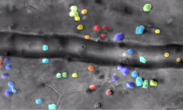
Captured by the lab of Jesse Schallek, assistant professor of ophthalmology and neuroscience, the image shows microscopic immune cells escaping a nearby blood vessel in response to inflammation. The color overlay shows computer detection of single cells that are tracked over time. (Courtesy of Schallek lab)
Rochester researchers demonstrate way to track the interactions of microscopic immune cells in a living eye without dyes or damage, a first for imaging science.
Combining infrared videography and artificial intelligence, the new technique could be a 'game-changer' for some clinical diagnoses as well as for fields like pharmaceuticals.
University of Rochester vision scientist Jesse Schallek can barely contain his excitement as he shares time-lapse videos showing immune cells moving through living retinal tissue at the back of an eye.
In one clip, immune cells crawl so slowly along the inside edge of a blood vessel that the video must be sped up 25 times to show their progress. Another cell slowly treads against the flow of blood in a vessel, like a salmon fighting its way upstream. Other immune cells leave the blood vessels and inch through the surrounding tissue, then congregate in a swarm, forming a beehive of activity.
Schallek and his vision lab at the University of Rochester Center for Visual Science and Flaum Eye Institute, have created a new microscopy technique, described in the journal eLIFE, that builds upon groundbreaking adaptive optics developed at the University more than 20 years ago.
Combined with time lapse videography and artificial intelligence software, the new technique enables researchers for the first time to noninvasively image and track—without labeling—the interactions of translucent immune cells within live retinal tissue in animals. Until now, the immune cells had to be labeled with fluorescent agents and often reinjected in order to image them—raising questions about how this might change the behavior of the cells. Another common, but limiting approach is to remove cells and study them with a microscope in a dish.