menu

News

20242023202220212020

University Research Award helps team explore regeneration in a critical layer of the cornea

Friday, December 9, 2016

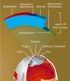
Illustration of structure of cornea

The structure of the cornea.
(Keratomania.com eye diagram by
Chabacano,via Wikimedia Commons.)

On the backside of the cornea is a single layer of cells that plays an all-important role, maintaining just the right fluid balance to keep the cornea transparent so that light can enter the eye. Until recently, it was believed this layer, called the corneal endothelium, is incapable of replacing its damaged cells. As more cells become damaged, the cornea becomes opaque, leading to loss of vision and, ultimately, to as many as 30,000 endothelium transplants a year in the United States alone.

A team of University researchers is exploring the possibility that stem cells on the outer edges of the cornea, given the right stimulation, can migrate into the endothelium to replace damaged cells. (Undifferentiated stem cells develop into specialized cells.) The work raises the possibility of restoring vision without the need for transplants.

The team is led by Amy Kiernan, associate professor of ophthalmology, and includes Jannick Rolland, the Brian J. Thompson Professor of Optical Engineering; Patrice Tankam, a senior scientist in the Center for Visual Science; Changsik Yoon, a graduate student in Rolland's lab; Rebecca Rausch, a graduate student in Kiernan's lab; and Holly Hindman, former associate professor of ophthalmology, now in private practice but still consulting on the project. They are supported with a $75,000 University Research Award. The URA program is designed to help researchers develop preliminary data or proof of concept needed to leverage larger federal or foundation awards to carry a promising project to completion.

There have been tantalizing clinical hints that the corneal endothelium may have regenerative capabilities, Kiernan says. For example, there have been cases in which endothelial transplants failed to engraft, but the cornea cleared up anyway, with regeneration of the endothelium occurring on its own. "So it seems that if something is done that stimulates a progenitor or stem cell population, most likely those in the periphery of the cornea, there is some regenerative capacity in the endothelium -- just based on clinical studies," Kiernan says.

Her team will attempt to identify the potential stem cells that might be stimulated to migrate to the endothelium to repair damage. They will use mouse models from Kiernan's lab in which adult stem cells can be permanently tagged with fluorescent biomarkers and tracked even after they differentiate into other cells. The identification and tracking of those cells will be done by refining a novel imaging approach developed in Rolland's lab. Called Gabor domain optical coherence microscopy, the technology allows rapid, noninvasive imaging of cellular structures beneath the surface of the skin or within the human eye -- in greater detail than traditional imaging with optical coherence tomography.

"Think of it as a high-definition, volumetric imaging," Rolland says. "But we also want to know what kind of cells we are looking at, so we are integrating fluorescence imaging with the high-definition volumetric microscopy so we can do both." The team represents a combination of pertinent expertise: cell development and regeneration (Kiernan and Rausch), imaging (Rolland, Tankam, and Yoon), and the biological basis for corneal and ocular surface diseases in humans (Hindman). The University Research Award funding is helping support graduate students and technicians working on the project, and the cost of mice and supplies. "Pilot funding like this is so important, especially with NIH grants shrinking," Kiernan says.

"It's really helpful to be able to bridge this kind of interdisciplinary effort," says Rolland. "You need to work together a little bit to understand the challenges involved and what you need to do to secure preliminary data, to show we have a pathway. "It takes time to get data, so even a small grant that provides a bridge for a year or two can make a huge difference."

Brain training video games help low-vision kids see better

Monday, November 28, 2016

Studies going back several years have shown that playing action video games (AVG) can help improve visual acuity. A new study by vision scientists at the University of Rochester and Vanderbilt University found that children with poor vision see vast improvement in their peripheral vision after only eight hours of training via kid-friendly video games. Most surprising to the scientists was the range of visual gains the children made, and that the gains were quickly acquired and stable when tested a year later.

"Children who have profound visual deficits often expend a disproportionate amount of effort trying to see straight ahead, and as a consequence they neglect their peripheral vision," said Duje Tadin, associate professor of brain and cognitive sciences at Rochester. "This is problematic because visual periphery—which plays a critical role in mobility and other key visual functions—is often less affected by visual impairments."

"We know that action video games (AVG) can improve visual perception, so we isolated the AVG components that we thought would have the strongest effect on perception and devised a kid-friendly game that compels players to pay attention to the entire visual field, not just where their vision is most impaired," said Tadin, who is also a professor in the Center for Visual Science. "As a result, we've seen up to 50 percent improvement in visual perception tasks."

Read More: Brain training video games help low-vision kids see better

UR Medicine Flaum Eye Institute Welcomes Visionary Eye Associates

Wednesday, November 2, 2016

UR Medicine has acquired Visionary Eye Associates to join Flaum Eye Institute, a national leader in vision care and research.

The optometric practice of Michael D. DePaolis, O.D., F.A.A.O., and Robert A. Ryan, O.D., F.A.A.O., will continue to provide patients state-of-the-art vision care augmented by convenient access Flaum Eye Institute's specialized medical and surgical eye care

"Visionary Eye Associates is a well-respected team and they have a similar philosophy in how to deliver high-quality care," said Steven Feldon, M.D., M.B.A., director of the Flaum Eye Institute and chair of Ophthalmology at the University of Rochester Medical Center. "We are excited to have them join our team and expand opportunities for people to access Flaum Eye Institute services."

For nearly two decades, DePaolis and Ryan have served as clinical associates of Ophthalmology at University of Rochester School of Medicine and Dentistry. They have been appointed associate professors of clinical Ophthalmology, pending approval by the University of Rochester Board of Trustees.

"We are eager to be a part of URMC to serve more people and more closely collaborate with local primary care physicians to ensure seamless care," said Ryan, who started the practice with DePaolis in 1995. Their offices are in Brighton and Webster.

The Flaum Eye Institute is among the nation's leaders in vision care and research. Ophthalmologic specialists provide diagnostic testing, advanced surgical interventions and optimized management of the most complex eye conditions. Patients from around the world travel to Flaum for expert care for artificial cornea transplants, refractive surgical correction, retinal degenerative diseases management and treatment of orbital inflammatory processes. Scientists pioneer new diagnostics and treatments and are dedicated to translating their findings to clinical advances that preserve and enhance vision.

Read More: UR Medicine Flaum Eye Institute Welcomes Visionary Eye Associates

NGP Student Awarded NIH Fellowship

Monday, October 3, 2016

Rebecca Rausch, a fifth year neuroscience graduate student in Dr. Richard Libby's lab was awarded an NIH Individual Pre-doctoral Fellowship from the National Eye Institute for her project entitled: The Role of Notch and BMP Signaling in Anterior Segment Dysgenesis (2016-2019).

Congratulations Becca!

Play it Safe: Avoiding Common Eye Injuries

Tuesday, August 23, 2016

Child's play can be great fun, except when it results in an unexpected visit to the pediatrician or eye doctor. UR Medicine pediatric ophthalmologist Dr. Benjamin Hammond, of the Flaum Eye Institute, offers insight on some common childhood eye injuries.

Injuries can happen in the blink of an eye so use caution to help youngsters avoid corneal abrasions. These often result from:

Toy tinkering—The U.S. Consumer Product Safety Commission estimates about 250,000 children are taken to hospital emergency rooms for toy-related eye injuries each year. Injuries frequently occur because a child uses a toy that isn't appropriate for their age, or uses it in an unintended way. We've all seen a youngster swing a plastic doll or throw a heavy toy in anger, striking another child in the face. While any toy can be harmful if used inappropriately, extra care should be taken with toys used in simulated play-fighting or that launch projectiles, such as play swords or foam dart guns. These should never be swung or aimed towards the face. Many parents and children think these are harmless, but an unlucky hit or shot to the eye can cause injuries, including abrasions and bleeding inside the eye. Most heal without long-term impact on vision but they often need eye medications to prevent complications.

Balls and bats—Budding athletes face injuries on the field and in the backyard where opportunities for being struck in the face by sports balls, fingers, elbows or equipment are plentiful. A blunt force hit or poke in the eye can cause bleeding or retinal detachments, which require immediate medical attention. A child who complains of blurry or cloudy vision, light sensitivity or flashes should see a doctor to check for an internal injury to the eye. Well-fitting protective eyewear can prevent up to 90 percent of sports-related injuries. And though few sports require routine eye protection, children with poor vision, previous eye surgery, or quality vision in just one eye should wear it.

Pet problems—Eye doctors frequently care for young children who suffer a dog bite near the eye. The family dog may be well-trained, but pet behavior can be unpredictable. Many times a dog bite near the cheek or eye will damage tear ducts. Parents can help avoid injury by teaching youngsters how to interact safely with pets.

Supervised play is one of the best ways to help reduce risky situations as children grow and gain understanding of the potential dangers of their behavior.

Read More: Play it Safe: Avoiding Common Eye Injuries

URMC Refractive Surgeon Selected for International Award

Friday, July 15, 2016

Scott MacRae, M.D., director of the Refractive Surgery Center at the University of Rochester Medical Center's Flaum Eye Institute, was recently tapped to receive the Jose I. Barraquer Lecture and Award from the International Society of Refractive Surgeons (ISRS), a partner of the American Academy of Ophthalmology. The award, named after the father of refractive surgery, is a top honor for refractive surgeons who have made significant contributions to the field and who demonstrate dedication to advancing the science of refractive vision correction.

In over 25 years as a refractive surgeon, MacRae has performed more than 20,000 refractive procedures, trained over 400 refractive surgeons from around the world, and helped develop several new refractive surgery instruments and techniques -- including a personalized system for laser vision correction. He also holds the patent for the Rochester Nomogram, a complex system of measurement for visual abnormalities that immensely improved the success rate of custom laser vision correction.

Before joining the University of Rochester Medical Center in 2000, MacRae had 16 years of prior experience as a corneal specialist at Oregon Health Sciences University. There, he encountered several corneal transplant patients who had vision issues after their operation that could not be explained using conventional measurement systems. At the time and for 200 years prior, ophthalmologists could measure only three things: nearsightedness, farsightedness, and astigmatism -- all considered "low order aberrations".

Meanwhile, David Williams, Ph.D., dean for research of Arts, Sciences, & Engineering and William G. Allyn Professor of Medical Optics at URMC, changed that by developing the Zywave™ wavefront sensor with Bausch & Lomb. Wavefront sensor technology could measure a whole spate of corneal and lens irregularities that were previously virtually undetectable. These "higher order aberrations" were the cause of the post-operative vision issues in MacRae's corneal transplant patients.

Read More: URMC Refractive Surgeon Selected for International Award

Proposal by Amy Kiernan Receives University Research Award

Monday, May 23, 2016

A collaborative project involving Associate Professor Amy Kiernan of the Flaum Eye Institute has been chosen as one of the 2016-17 University Research Awards. One of just eight applications chosen by senior research leadership, the proposal entitled, "Understanding cell turnover and injury recovery in the corneal endothelium" will be funded $75.000 annually.

Congratulations to NGP student Aleta Steevens

Friday, April 8, 2016

Photo of Aleta Steevens

Aleta Stevens, an NGP student in Dr. Amy Kiernan's lab, secured a 3-year NIH Individual Pre-doctoral Fellowship, F31 entitled, "Elucidating the role of SOX2 in inner ear development."

Excellent work Aleta!

Neuroscience Graduate Students Win Award for Teaching

Friday, April 8, 2016

Aleta Steevens

Neuroscience Graduate Program students, Aleta Steevens (Dr. Amy Kiernan lab) and Heather Natola (Dr. Chris Pröschel lab) were awarded the 2016 Edward Peck Curtis Award for Excellence for Graduate Student Teaching.

Only a handful of these are awarded each year, and all this year's nominees were extremely well-qualified.

Congratulations to both!!!

ABVI Honors Flaum Eye Institute Leader for Dedication to Community

Monday, March 28, 2016

UR Medicine Flaum Eye Institute Director Steven Feldon, M.D., M.B.A., will be honored by the Association for Blind and Visually Impaired during its 2016 Visionary Gala: Sea Great Things Saturday, April 9.

"Dr. Steven Feldon's support of, and interest in, the work of ABVI are truly distinguished and genuinely appreciated," said Gidget Hopf, ABVI's president and CEO. "His passion for ABVI's work led him to become a board member. While on the board, he helped develop and grow our person-centered programs and services. It is for these reasons we honor Dr. Feldon this year."

A neuro-ophthalmologist and orbital surgeon, Feldon joined University of Rochester Medical Center in 2001 as chair of the Department of Ophthalmology, and is founding director of the growing David and Ilene Flaum Eye Institute.

He has overseen the growth of his department from a faculty of five clinicians and three basic scientists to its current complement of 23 clinicians and eight basic scientists. Over the last decade, the number of National Institutes of Health grants has risen to 10 and there are now has four clinical care locations in Rochester and Geneva.

"I am very pleased to receive this honor from ABVI-Goodwill of the Finger Lakes," Feldon said. "I was privileged to serve for nine years on the Board, during which time ABVI made tremendous strides in empowering those with visual challenges through rehabilitation, community service, and job creation. The organization is well-served by the tireless, enlightened leadership of Gidget Hopf, her board of directors, and dedicated staff.

"The Flaum Eye Institute has partnered with ABVI to provide eye care for the underserved, vision screenings and research in visual rehabilitation. We work together to support people who undergo intraocular telescope implantation for management of vision loss due to macular degeneration. We live in a wonderful community enhanced tremendously by agencies such as ABVI-Goodwill."

Read More: ABVI Honors Flaum Eye Institute Leader for Dedication to Community

Doing something larger than you could ever do on your own

Friday, February 12, 2016

"There is a tendency for many investigators, especially early in their careers, to hold onto their work and not share it," says David Williams, the William G. Allyn Professor of Medical Optics; Dean for Research in Arts, Sciences and Engineering; Director of the Center for Visual Science - and a leading eye expert who pioneered the use of adaptive optics for vision correction.

"They don't realize - and it's one of the things that took me longer to learn than I wish it had - that one of the best ways to build your reputation is to share your ideas or your technology with the hope that they will be adopted.

"I was lucky enough to realize that if I let my students take my adaptive optics technology and use it to build their own labs, for example, it not only helped them get their independent research programs off the mark but also enhanced my reputation because so many more people were able to access and deploy the technology."

Is it any wonder then, that of the five NEI Audacious Goals grants recently awarded to Williams and four other investigators:

  • four of the projects use adaptive optics as their core technology?
  • three of the other PI's are either current collaborators with Williams or former postdocs in his lab?
  • which means that four of the PI's will be cooperating with each other, even as they individually collaborate with other experts in the field on their individual projects - in effect widening the opportunities for synergy?

    "That's the excitement of this," Williams says. "Why should we compete when one group can do one piece of it, and a second group can do another, and as along as you can manage authorships and credit appropriately and fairly, we can be much more efficient and effective in getting things done?"

    "One of the things I'm proudest about in this community of people around the world doing adaptive optics and retinal imaging is that almost all of us get along really well, and we're moving science forward as rapidly as we can by helping each other. That doesn't always happen in science."

    As Dean of Research for Arts, Science and Engineering, Williams is always looking for young faculty throughout AS&E who have the right personality and vision to take on larger, multi-investigator, multi-institutional projects.

    "You have to be gregarious and interested in working with other people and tolerating the quirks that they have, just as they have to tolerate the quirks you have," Williams said.

    "The largest source of optimism for me about the AS&E research portfolio is the quality of our junior faculty members - their enthusiasm and energy. Many of them have cut their teeth on individual investigator awards and will reach a certain point in mid career when they realize they need to reach out for complementary expertise in order to do more."

    Williams' advice: The best collaborator may not be the first one that comes to mind.

    "One of the biggest mistakes faculty members make is to choose a collaborator who is just like them, who has the same interests in a problem and the same background and who they can easily begin a conversation with because they are so closely aligned. But that doesn't really help your research. You want to have somebody who . . . has a completely different skills set. As obvious as that is, it doesn't always get factored into planning how to accumulate the necessary wisdom to do something larger than you could ever do on your own."

Talented Students Raise Funds to Help City Pupils Who Need Eyeglasses

Friday, February 12, 2016

UR Medicine's Flaum Eye Institute received a $20,000 donation to support its program to provide free vision care for needy schoolchildren. Kids Reaching Hearts Through Performing Arts, an organization of area high school students dedicated to supporting children through artistic expression, raised the funds during a gala event and variety show in October.

"We are grateful to see such talented students dedicated to helping others," said Steven Feldon, M.D., M.B.A., director of the Flaum Eye Institute and chair of Ophthalmology at the University of Rochester Medical Center. "It saddens us to know that children go to school and are not able to perform to the best of their ability because of a vision problem. These funds will help children succeed."

The funds will be used to support the Glover-Crask Charitable Trust Eyeglasses for Kids program, providing vision exams and eyeglasses to children who attend schools in Rochester and Finger Lakes region. Ophthalmologists and support staff work closely with principals and school health leaders to identify youngsters who need eye exams, and if they need eyeglasses, the FEI team crafts them immediately.

"It's heartwarming to see the smiles on the kids' faces when they receive the glasses and to know how much it means to them to go home seeing better," Feldon said. The endowed program began a year ago with funding from the Glover-Crask Charitable Trust, led by John Harris, an FEI Board member, and has grown quickly.

Read More: Talented Students Raise Funds to Help City Pupils Who Need Eyeglasses