URMC / Labs / Romanski Lab / Projects / Connections of Auditory and Visual Ventral Prefrontal Neuron
Connections of Auditory and Visual Ventral Prefrontal Neurons
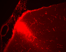
Fluorescent tracer injection in the prefrontal cortex
We hypothesize that although most (and perhaps all) of the VLPFC is multimodal, that there are still tendencies towards dominant responses in the VLPFC with predominantly auditory neurons localized anterior to visual object and face-specific neurons. Though our recent physiology suggests this but it has not been fully tested. Secondly, our current anatomical tracing studies (Diehl et al., 2007) indicate that VLPFC neurons receive afferents from unimodal auditory and visual regions and from polymodal cortical regions.
Innervation of ventrolateral prefrontal cortex from anterior auditory belt association cortex. (Romanski et al., 1999)
To determine the fine detail of this organization which has great implications for the domain specificity hypothesis we combine anatomy and physiology and place tracers in physiologically mapped regions of auditory, visual and multimodal regions of VLPFC. IN previous experiments we have recorded auditory, visual, and multisensory responsive neurons from VLPFC and using these physiologically-defined boundaries, placed anatomical tracers into distinct locations to determine the afferent and efferent projections of the auditory and visual responsive cells. Our results demonstrate that prefrontal auditory neurons receive the densest input from areas within the superior temporal gyrus (STG), and to a lesser extent TPO.
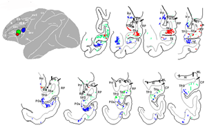
Retrograde labeling from injections of tracer into auditory (red), visual (blue) and multisensory (green) responsive regions of the ventrolateral prefrontal cortex
In contrast, visually responsive neurons in VLPFC receive afferent projections from a number of inferotemporal cortex areas including TE, IPa, PGA and TPO. Injections placed in the inferior convexity between the auditory and visual selective regions, where neurons responded to both auditory and visual stimuli received inputs mainly from TPO and to a lesser degree areas TAa, IPa and TE. By investigating the anatomical connections of the VLPFC, we hope to better understand how auditory and visual information reaches the frontal lobes.
« back to all projects