URMC / Labs / Wismüller Lab / Projects / Soft Tissue Characterization of Human Patellar Cartilage
Soft Tissue Characterization of Human Patellar Cartilage
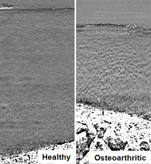
Human patellar cartilage from healthy (left) and
osteoarthritic (right) specimens, as visualized on PCI-CT.
Phase contrast X-Ray Computed Tomography (PCI-CT) is a novel imaging technique that provides soft-tissue discrimination in cartilage tissue at a micrometer scale resolution. We are currently investigating the use of texture features that characterize properties of the cartilage matrix (visualized with PCI-CT) as imaging bio-markers for tracking osteoarthritis progression.
We specifically focus on chondrocyte patterns in the radial zone of the cartilage matrix. Previous research has shown that chondrocytes are aligned in so-called Benninghoff arches radiating from the tide mark in healthy cartilage. Osteoarthritic cartilage exhibits a more disorganized clustering of chondrocytes in the radial zone. We use texture features derived from statistical, topological and geometrical approaches to capture such differences in patterns observed in healthy and osteoarthritic cartilage specimens.
We currently focus on using such texture features for classification of regions of interest (ROI) extracted from cartilage ex vivo specimens as being healthy or osteoarthritic. We would like to extend the use of such features to a multi-class classification problem where we differentiate between ROIs exhibiting patterns indicative of different stages of osteoarthritis.
Journal Publications
-
M.B. Nagarajan, P. Coan, M.B. Huber, P.C. Diemoz, C. Glaser and A. Wismüller, “Computer-Aided Diagnosis for Phase Contrast X-ray Computed Tomography: Quantitative Characterization of Human Patella Cartilage with High-Dimensional Geometric Features,” Journal of Digital Imaging 27(1):98-107, (2014). PMID: 24043594
-
M.B. Nagarajan, P. Coan, M.B. Huber, P.C. Diemoz, C. Glaser and A. Wismüller, "Computer-Aided Diagnosis in Phase Contrast Imaging X-ray Computed Tomography for Quantitative Characterization of ex vivo Human Patellar Cartilage," IEEE Transactions on Biomedical Engineering 60(10):2896-2903 (2013). PMID: 23744660
Conference Publications
-
M.B. Nagarajan, P. Coan, M.B. Huber, P.C. Diemoz, and A. Wismüller, "Phase contrast imaging X-ray computed tomography: quantitative characterization of human patellar cartilage matrix with topological and geometrical features," Proceedings of SPIE Medical Imaging 9038:111-118 (2014).
-
M.B. Nagarajan, P. Coan, M.B. Huber, P.C. Diemoz, C. Glaser and A. Wismüller, "Characterizing healthy and osteoarthritic knee cartilage on phase contrast CT with geometric texture features," Proceedings of SPIE Medical Imaging 8672:1J1-1J8 (2013).
-
M.B. Nagarajan, P. Coan, M.B. Huber, C. Yang, C. Glaser, M.F. Reiser, and A. Wismüller, "Characterization of healthy and osteoarthritic chondrocyte cell patterns on phase contrast CT images of the knee cartilage matrix," Proceedings of SPIE Medical Imaging 8317:201-208 (2012).
« back to all projects