Visualization of Viral Particles by Negative Staining Electron Microscopy
Electron microscopy is a useful tool to examine virus morphology. A negative staining technique uses heavy metal salts to enhance the contrast between the background and the virion's image. This is a very simple and direct technique. Pelleted viral particles are resuspended in distilled water. A drop of viral suspension is placed on a petri dish. A Formvar-coated EM grid is placed Formvar side down on top of the virus drop for approximately 1-3 minutes. The grid is removed, blotted with filter paper and placed onto a drop of 2.0% phosphotungstic acid (PTA), pH 7.0, for one minute. The excess PTA is removed, and the EM grid can now be examined and photographed for EM.
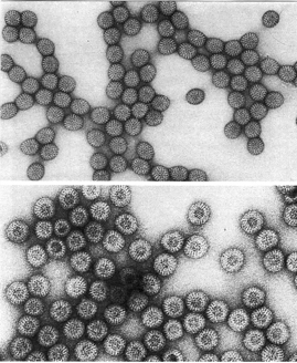
Negative stain of rotavirus (Ref: Palmer, E.L., Martin, M.L.: Electron Microscopy in Viral Diagnosis, pg. 77, CRC Press, Inc., 1988.)
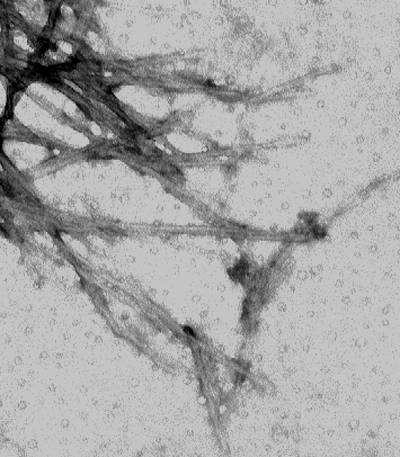
Negative stain of alpha synuclein (Dr. Howard Federoff's research group)