Routine Processing
Tissue is fixed in EM fixative (2.5% glutaraldehyde or 4.0% paraformaldehyde/1.0% glutaraldehyde) that we can provide. The tissue is rinsed in buffer, post-fixed in 1.0% osmium tetroxide (OsO4) to preserve lipid and lipoprotein structures. The specimen is then processed through a graded series of alcohols, infiltrated in Spurr epoxy resin and embedded. The resin embedded tissue is polymerized at 70°C overnight. Semi-thin (1.0-2.0 um) sections are cut with a glass knife to select an appropriate area for ultra-thin sectioning with a diamond knife. Sections are placed on grids, stained with uranyl acetate and lead citrate, and examined by EM and photographed.
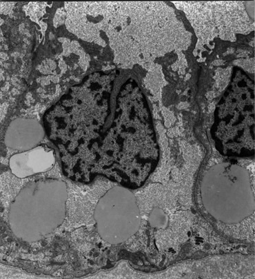
Electron micrograph of a renal cell carcinoma
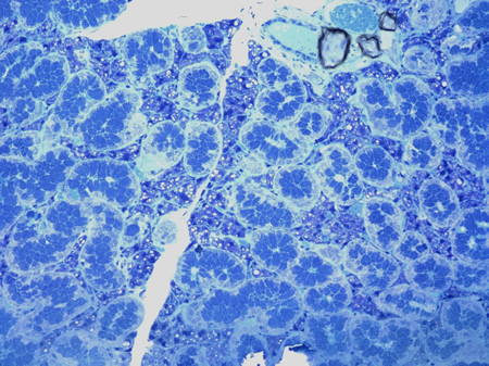
Toluidine Blue stained plastic 1micron section of a salivary gland