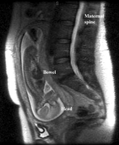
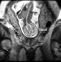
MR Imaging of the Fetus
MR imaging is occasionally done of the fetus. Usually it is to look for congenial abnormalities of the brain and often to clarify something seen on ultrasound. Below are examples of fetal MR images.
MR imaging complements ultrasound when additional information is needed to make treatment decisions during pregnancy. Up until recently, studies were limited by fetal motion but now the scanners are so fast that it is possible to get good images even if the fetus moves. Due to fast imaging techniques now available, images can be obtained in less than 1/2 a second. This means that neither the mother nor the fetus needs to be sedated. MR imaging has proven to be especially beneficial in evaluation of fetal brain abnormalities such as:
- Encephalocele see AJR 1999;172:813-818
- Agenesis of the corpus callosum
- Porencephaly - destruction of brain tissue
- Partial absence of the septi pellucidi
There is no known risk involving fetal MRI.