Neuron-Neuroglia Interactions
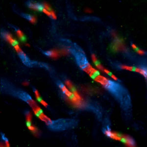
Optic nerve. Sodium channels (green) Caspr (red) potassium channels (blue)
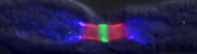
Propagation of electrical signals with high speed and reliability in nerve fibers depends on a complex interaction between neurons and their associated myelinating glial cells. As a result of this interaction ion channels are clustered at specific sites along the axon. This system is studied in both development and disease. At birth axons have little myelin, but by the end of the first postnatal week both glial ensheathment and ion channel clustering are at an advanced state. In multiple sclerosis myelin is damaged, and ion channel distributions are disrupted. The immune mechanisms responsible for this pathology are not known. This laboratory studies the molecular mechanisms responsible for these phenomena using electrophysiology, immunocytochemistry, molecular biology, and transgenic manipulations.
During myelination glial cells have a profound influence on the axonal ion channel distribution. In the peripheral nervous system Schwann cells bind to axons during both development and remyelination. Sodium channels, responsible for the upstroke of the action potential, cluster in the axon membrane adjacent to the tips of Schwann cell processes that are extended during early stages of ensheathment. As Schwann cells grow longitudinally, these channels appear to move with them, ultimately fusing with a neighboring cluster to form a node of Ranvier. Voltage-dependent potassium channels have a reciprocal relationship with sodium channels, and are located primarily in clusters several micrometers on either side of the node. New studies are focused on the central nervous system and combine immunocytochemistry with electrophysiology to determine the distribution and role of ion channels during development of the optic nerve. We seek to determine the cellular and molecular events responsible for ion channel localization and stabilization in the neuronal membrane.

Peter G. Shrager, Ph.D.
Principal Investigator


- Deletion of murine Sarm1 results in a microenvironment that delays peripheral nerve regeneration after injury.; Science translational medicine; Vol 17(819), pp. eadp9155. 2025 Oct 08.
- Neuronal protein phosphatase 1β regulates glutamate release, cortical myelination, node of Ranvier formation, and action potential propagation in the optic nerve.; bioRxiv : the preprint server for biology. 2024 May 10.
- Sarm1 is not necessary for activation of neuron-intrinsic growth programs yet required for the Schwann cell repair response and peripheral nerve regeneration.; bioRxiv : the preprint server for biology. 2024 Mar 08.
- Nuclear Inhibitor of Protein Phosphatase 1 (NIPP1) Regulates CNS Tau Phosphorylation and Myelination During Development.; Molecular neurobiology. 2022 Jan 06.