Case of the Month: Chronic Diarrhea
By Phoenix D. Bell, MD, MS and Mark G. Ettel, MD
Clinical History
A 56-year-old female with a past medical history of hypothyroidism, anxiety, and depression presents with chronic diarrhea and is referred for colonoscopy.
Recent History
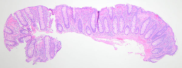
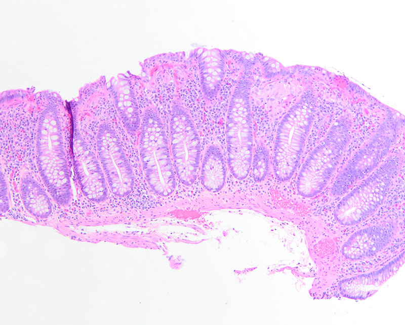
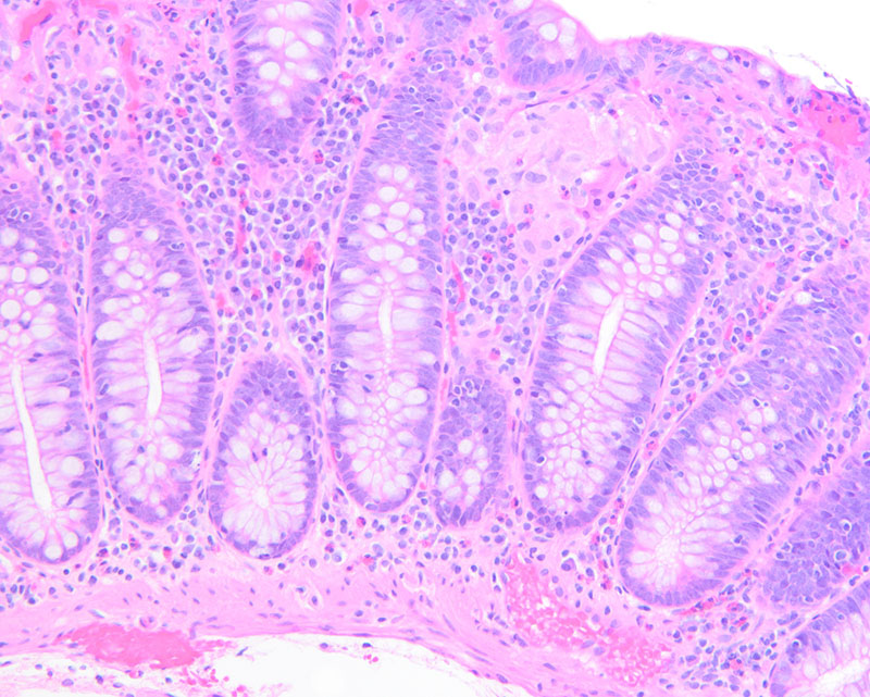
Colonoscopy reveals unremarkable colonic mucosa throughout the length of the colon. A random biopsy is taken and sent for histopathologic analysis. At low power, the crypt architecture is maintained and there is increased cellularity of the lamina propria (Figure 1). At medium power, the surface epithelium shows mucin loss and increased intraepithelial lymphocytes (Figure 2). Notably, there are collections of cells with abundant eosinophilic cytoplasm and multiple bland nuclei randomly distributed throughout the lamina propria below the surface epithelium (Figure 3). Acid-fast bacteria (AFB) and Gomori methenamine silver (GMS) stains are negative for microorganisms.