Case of the Month: Incidental Pancreatic Tumor
By Numbereye Numbere, MBBS (PGY-3) and Mark G. Ettel, MD
Clinical History
An asymptomatic middle-aged woman with an incidentally discovered pancreatic mass.
Past Medical History
The patient has a history of multiple endocrine neoplasia type 1 (MEN1), with a history of primary hyperparathyroidism, pituitary adenoma, mild hypercalcemia, and multiple lipomas.
Recent History
Abdominal CT showed a hypodense lesion in the tail of the pancreas. A follow-up MRI confirmed a 2.4 cm pancreatic mass and incidental bilateral non-obstructing renal calculi. Distal pancreatectomy was performed.
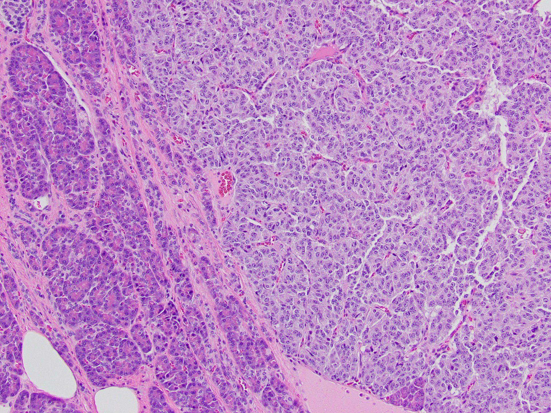
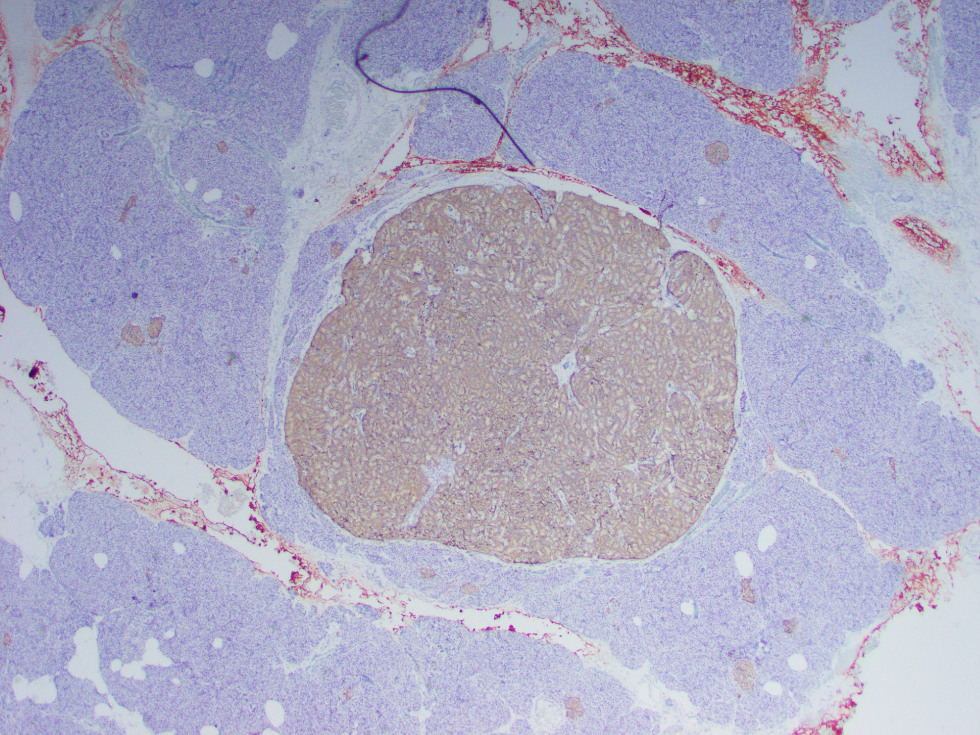
Gross examination of the distal pancreatectomy and splenectomy specimen revealed a 2.6 cm firm, well-circumscribed, homogeneous, yellow/tan intrapancreatic tumor and an unremarkable spleen.
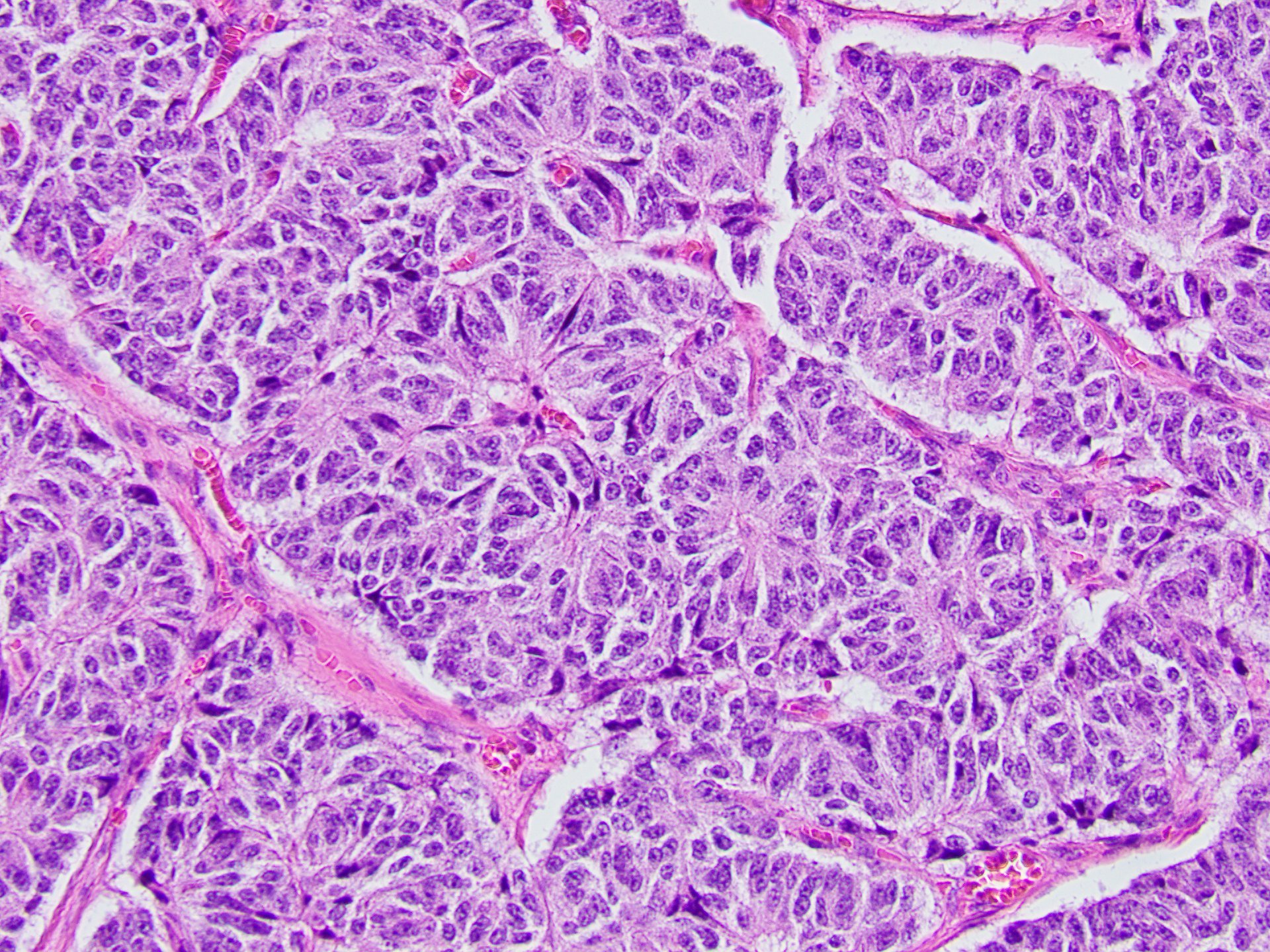
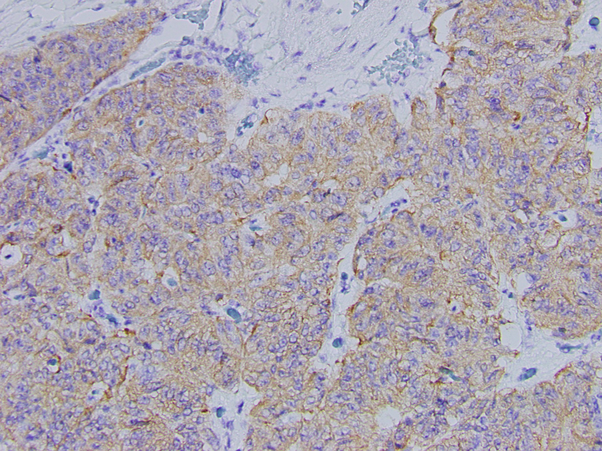
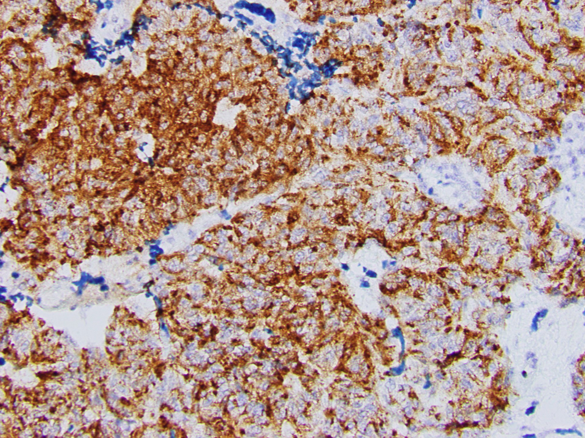
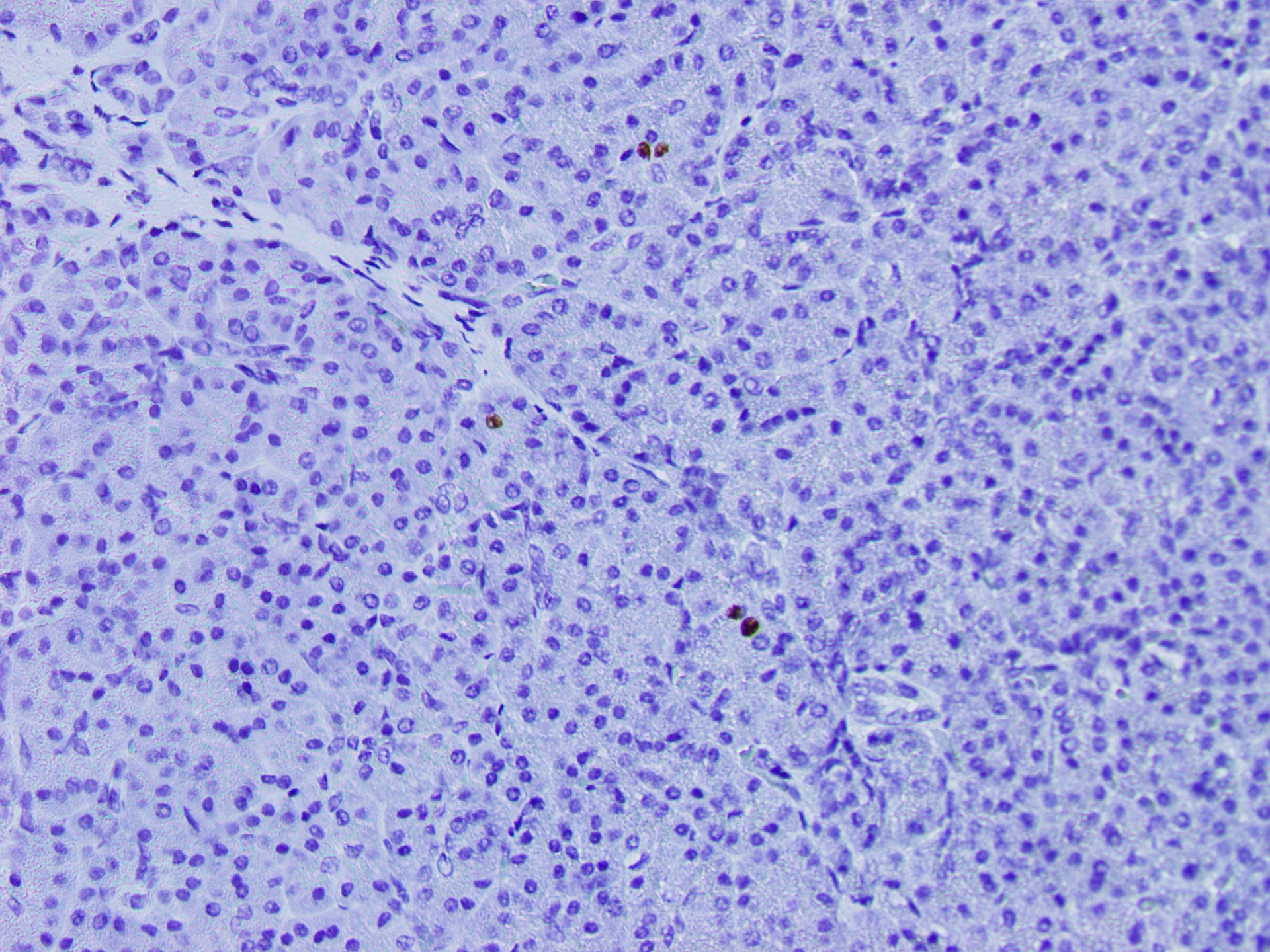
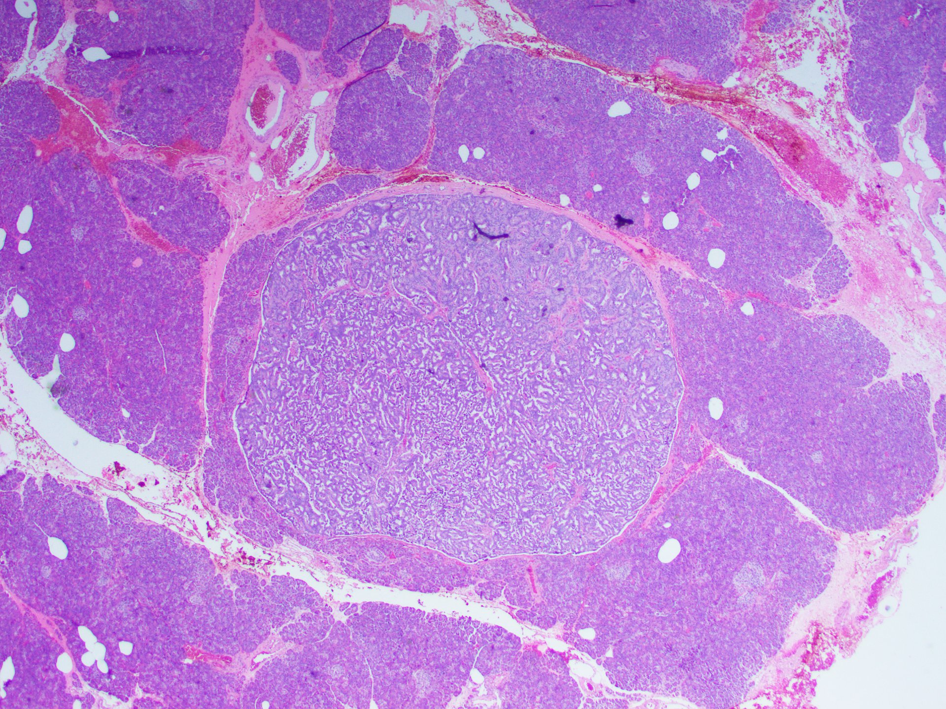
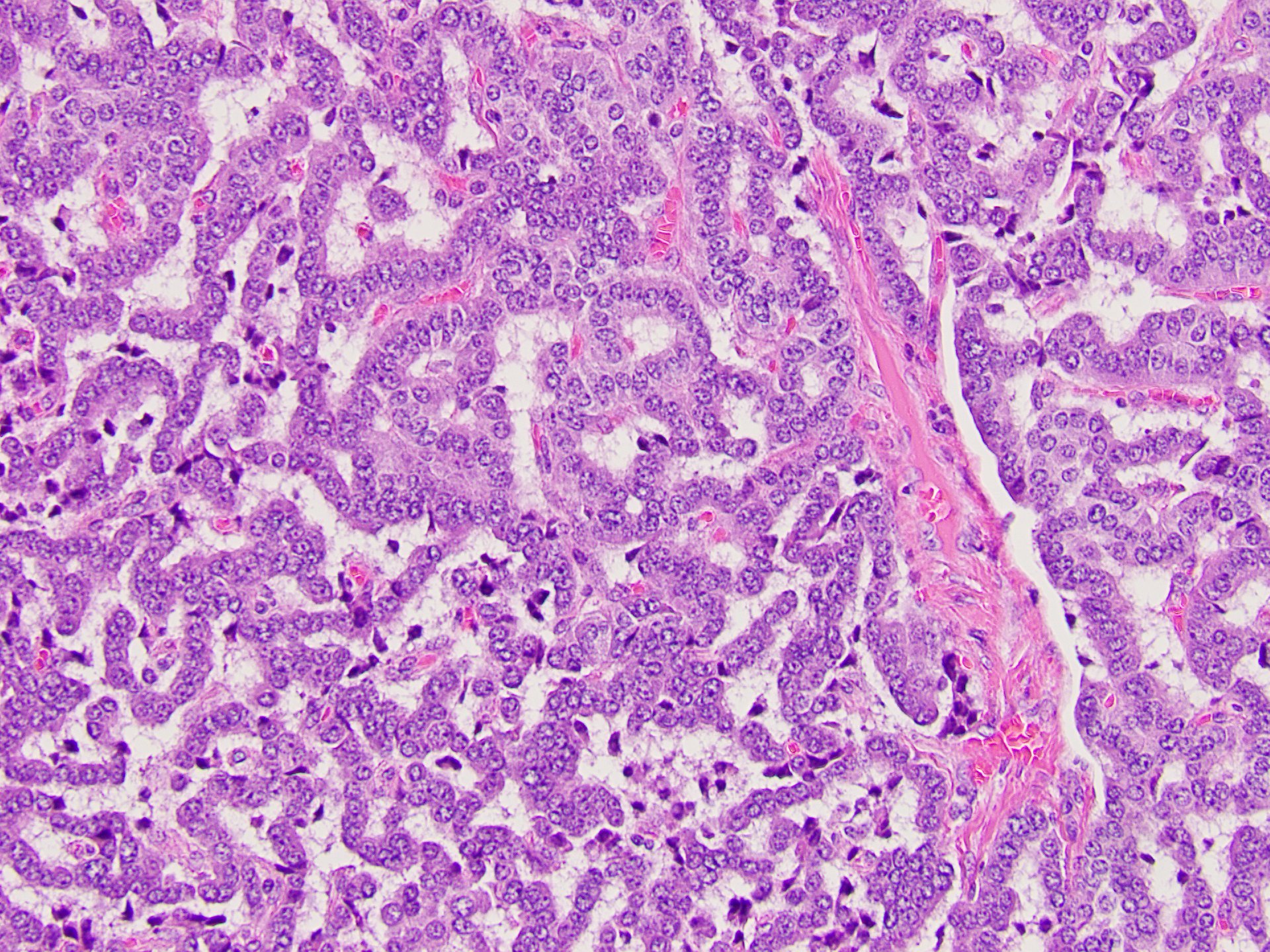
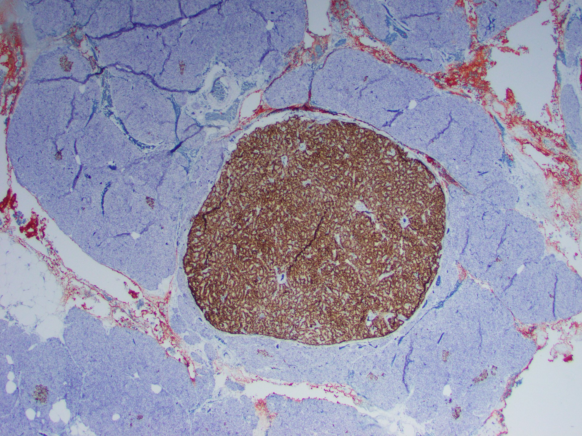
On microscopy, the tumor consisted of a monotonous population of round cells with dispersed fine chromatin, prominent central nucleoli, and a moderate amount of cytoplasm (Figures 1-5). Also, multiple small neuroendocrine lesions of similar appearance (microadenomas; Figures 6-9) were seen.