Unintentional Weight Loss and Abdominal Pain
Case Authors: Juwairiya Arshi, MBBS; Arash Lahouti, MD
Clinical History
A 75-year-old female presented with unintentional weight loss and abdominal pain.
Imaging
CT scan of the abdomen showed a heterogeneous retroperitoneal mass measuring 8.3 cm. CT scan of the chest revealed numerous bilateral round pulmonary nodules and an enlarged paratracheal lymph node.
Recent History
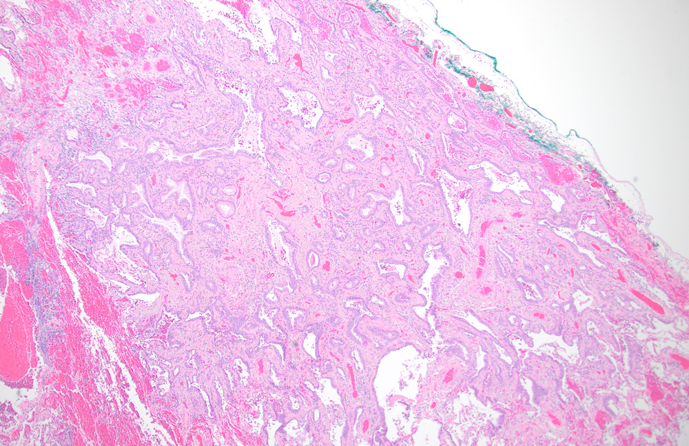
She underwent wedge resection of the right lower lobe. The wedge resection showed a highly pleomorphic malignant neoplasm (not discussed here) and an incidental nodule, which was not seen on gross examination.
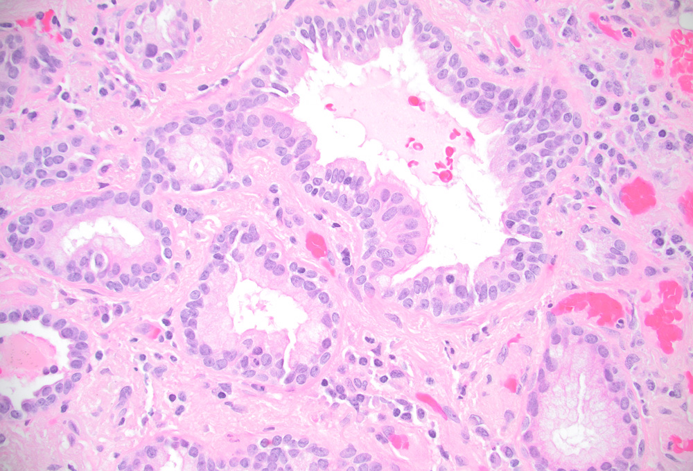
Histologic examination of this nodule revealed a subpleural 0.4 cm tumor (Figure 1). The tumor was composed of glands covered by bilayered (luminal and basal) epithelium. The luminal cells were comprised of a mixture of ciliated columnar cells and mucus cells (Figure 2).
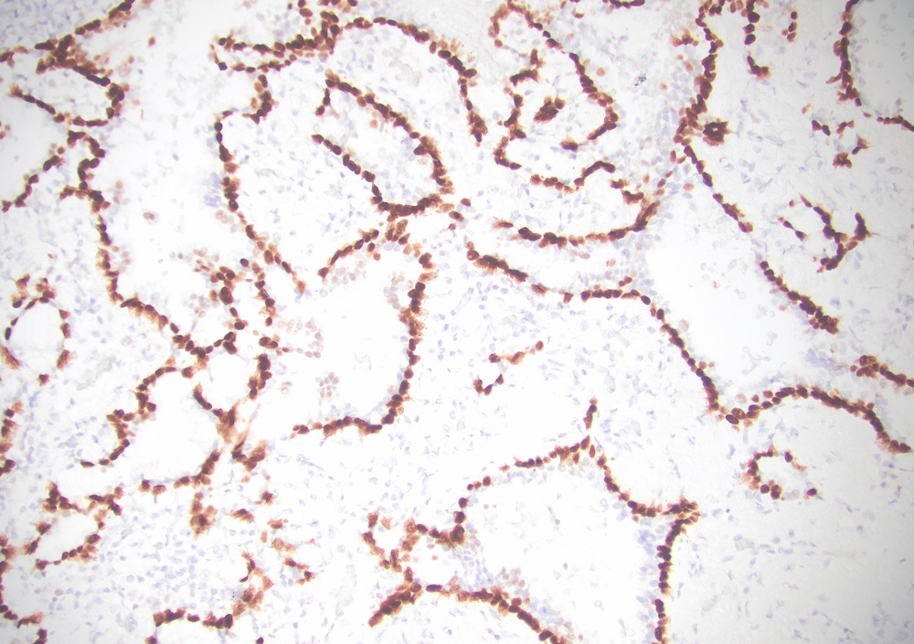
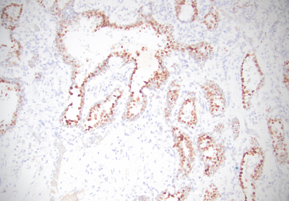
Immunohistochemical examination of the tumor cells showed nuclear expression of TTF-1, and p63 in luminal and basal cells, respectively.