


Why Learners Choose Us
Why Learners Choose Us
The School of Medicine and Dentistry’s integration with University of Rochester Medicine, a leading academic health system within a top-tier research university, affords opportunities to form collaborative partnerships with researchers and practitioners from multiple academic and health care disciplines.
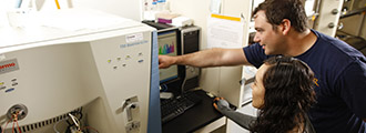
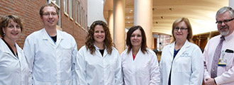
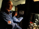
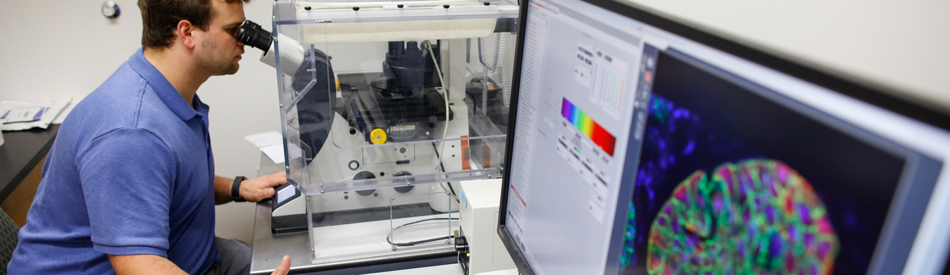
Individualized attention, close working relationships with your peers and faculty, and extensive participation in research are what you can expect from your experience in our graduate programs. We are a welcoming academic community with faculty mentoring, interdisciplinary programs, and progressively independent research opportunities. Our collaborative environment allows you to pursue intellectual challenges in your specific field, as well as in other related areas of interest.
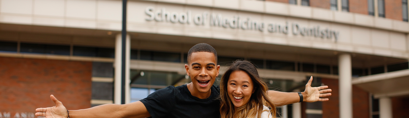
The School of Medicine and Dentistry offers more than 30 PhD, master’s, and advanced certificate programs encompassing the biomedical and health sciences.
As the fourth largest city in New York, Rochester is big enough to offer an array of cultural, recreational and leisure activities, and intimate enough to live affordably and navigate easily. With a cost of living significantly below the national average and proximity to the state’s beautiful Finger Lakes region, many who come here to train decide to stay. Discover all that Rochester has to offer.