Laser Lesion Model
Tracking the immune response to deep retinal lesions
 AOSLO provides a versatile platform for studying immune cell behavior in the mouse retina. One focus in our lab is to study immune cell interactions within the living retina. Here we induce outer retinal laser damage with 488 light, providing a sterile inflammatory microenvironment whereby we can track immune cell behavior.
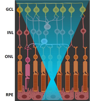
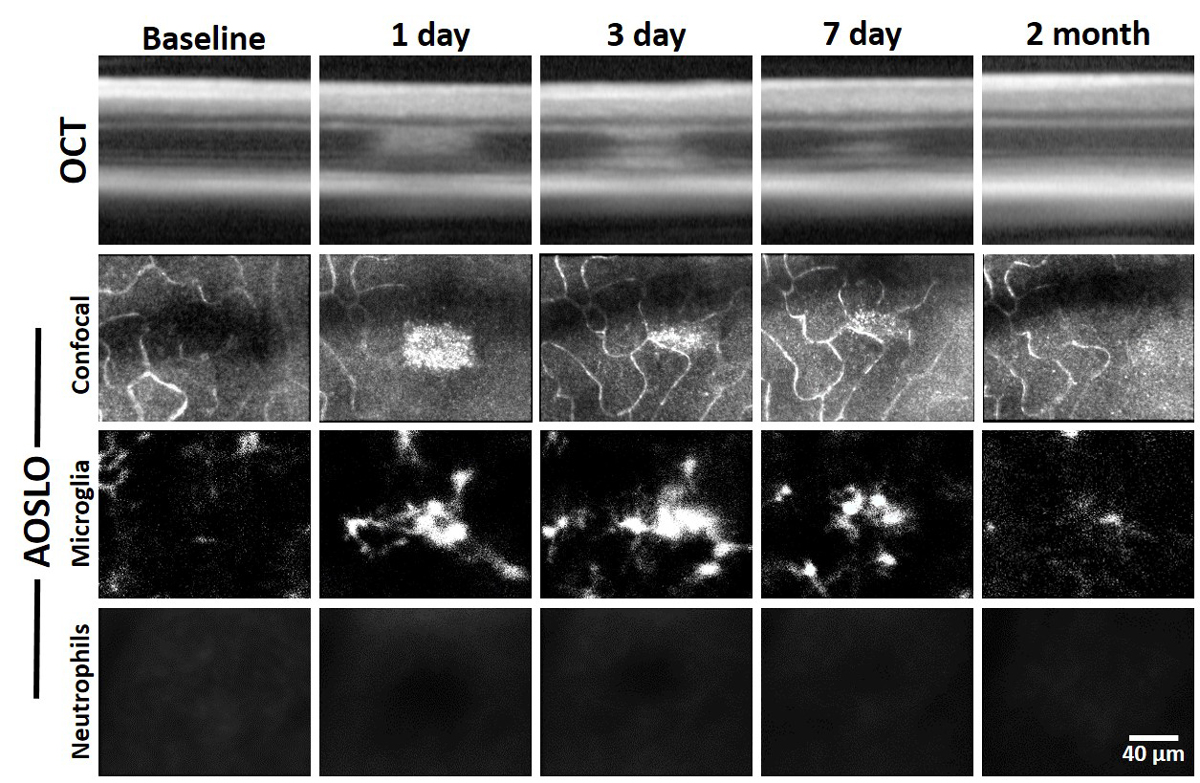
AOSLO provides a tool whereby we can both apply focal lesions to the outer retina AND non-invasively study the temporal dynamics of the immune cell response. We are interested in investigating the interaction between retina-resident microglia and systemic neutrophils. With optical coherence tomography and confocal AOSLO, changes in the reflectance profile of the retina are revealed. Here, we note a hyperreflective band centered at the outer nuclear layer by 1 day that is mostly resolved by 2 months. AOSLO also allows for simultaneous fluorescence imaging. Here we see that microglia swarm toward the focal damage and neutrophils are not present.
This work was presented at ARVO 2023.
Time-lapse fluorescence AOSLO allows us to track single neutrophils over time. Here we show that despite the robust microglial response, neutrophils do not extravasate into the retinal parenchyma and can be documented flowing through the capillaries just above laser damaged sites.
In addition to in vivo imaging, our lab routinely performs ex vivo histology on fixed retinal tissue. This approach not only confirms in vivo findings captured with AOSLO, but reveals additional details about the immune response to outer retinal damage.
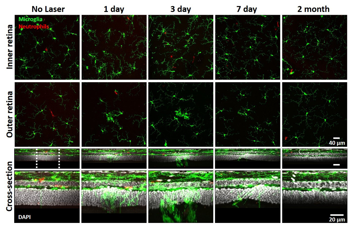
Ex vivo confocal microscopy provides enhanced resolution and sectioning capabilities. Here we reveal microglia phagocytosing photoreceptor somata.
With ex vivo confocal we can investigate morphological changes that immune cells undergo. Our lab also uses an experimental induced uveitis (EIU) model to induce systemic immune activation via intravitreal lipopolysaccharide (LPS) injection. Here we show neutrophils bound within capillaries are morphologically different than activated neutrophils.