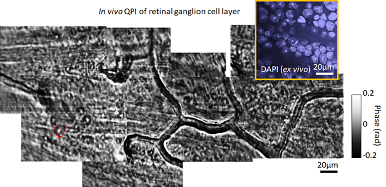
3D quantitative phase imaging for the single retinal cells in the living retina
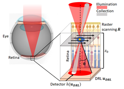
Most retinal cells are highly translucent by design. We have utilized phase-contrast adaptive optics scanning light ophthalmoscopy is an imaging tool that provides enhanced contrast for visualizing these cells in vivo. However, it is difficult to evaluate the actual cell morphology. In our lab, we have proposed an angle-resolved AOSLO (AR-AOSLO) to achieve 3D quantitative phase imaging (QPI) in the living retina with novel angle-space measurements. By imaging the forward scattered light projected on the deep reflective retinal layer (photoreceptor-RPE complex), the AR-AOSLO measures the angular spread of the light scattered by the cells.
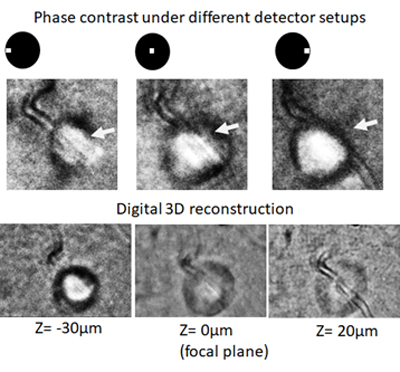
This strategy provides an additional image dimension to resolve the angular scattering behavior of the light as it propagates through translucent cells, which allows the recovery of the underlying optical properties of these cells, such as refractive index, phase, and axial positions. By solving the inverse problem of light scattering, QPI and 3D refractive index tomography can be realized computationally with AR-AOSLO, similar to computational microscopy. We have imaged the 3D phase for various in vivo retinal cellular structures, including leukocytes, photoreceptor somas, capillaries, and microglia.