Retinal vascular structure in the healthy lab mouse
In the mouse, there are very few studies aiming to provide a thorough analysis the retinal vascular network in health. Our lab is working solidify our knowledge within this field. Understanding of the retinal vasculature is important on multiple fronts. First, its baseline activity must adequately supply the metabolically demanding retinal tissue. Second, it is directly involved in neurovascular coupling processes that control the flow of blood to regions of high neural activity. Third, its structure, function, and integrity can all be altered in disease states such as diabetic retinopathy. Before we can understand the effect of disease on vascular components, first we need to provide a baseline in the healthy mouse.
Approach: Tracing the vascular tree
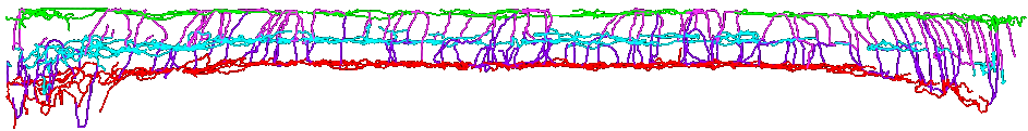
Our lab uses a confocal microscope to obtain high resolution z-stacks from ex vivo flat mounts extracted from the NG2-DsRed mouse. These mice express DsRed fluorescence within retinal vascular mural cells. ImageJ plugin Neuroanatomy SNT was used to trace all vascular paths in 3-D cubes. With this approach, we have shown that the mouse has a robust trilaminar structure that corresponds with the retinal nerve fiber layer, the inner plexiform layer and the outer plexiform layer. The mouse eye is small (3 mm diameter) and within this tiny space, we find that on average, each eye contains a one-meter total path-length of retinal vasculature!
En-face projection:
Cross-section:
Researchers across the world can now compare vascular changes in response to retinal degeneration/disease to an established baseline/healthy condition.