Immune Cell Imaging
The retina, being a ‘window’ to the brain and the rest of the body, offers the opportunity to non-invasively study single cells of the immune system in their native micro-environment, and also inspect the global health of the body. Our lab’s work on immune cell imaging examines the highly dynamic immune function from milliseconds-to-months in conditions of health and disease.
- Using AOSLO to image immune cells in the living eye non invasively
- Imaging microglia dynamics in health and disease
- Imaging immune cell movement within retinal vessels as a biomarker for inflammation
Using AOSLO to image immune cells in the living eye non invasively
Conventionally, inflammation has been studied by enucleating the eye and tagging different subsets of immune cells with fluorescent markers and look under a microscope. However, most ex-vivo techniques provide a single snapshot of the immunological response and do not speak about its regional specificity.
In Schallek lab, we are interested in studying and identifying immune cell behaviour in vivo as an indicator of the inflammatory state of the retina. For this, we are using a technology called adaptive optics to image immune cells without any labels inside a living animal. There are three major ingredients to achieve this.
- Micron level resolution: It is challenging to see single immune cells in conventional imaging modalities used clinically due to lack of adequate resolution and contrast. With adaptive optics can measure and correct for the aberrations of the eye, to give us unprecedented resolution to see single cells in the retina. In the image below, an inflamed murine retina was imaged with conventional imaging modality available in the clinic. With adaptive optics, we attain the required resolution to image single cells.
- Spatial Contrast: Immune cells very translucent, so we have incorporated phase contrast technique based on offset aperture. This allows us to see the motility of these cells with better contrast than just a confocal approach. For more details about our work on the phase contrast modality, refer to: Guevara-Torres A, Williams DR, Schallek JB. Origin of cell contrast in offset aperture adaptive optics ophthalmoscopy. Opt Lett. 2020 Feb 15;45(4):840-843. doi: 10.1364/OL.382589. PMID: 32058484; PMCID: PMC7337096.
- Stable timelapse imaging: We need a stable platform to monitor and track immune cells over time, since their motility differentiates them from other cell types in the retina.
These 3 innovations allow us to study immune cells behaviour from the start of inflammation to its resolution.
With adaptive optics, we can image and measure different stages of immune cell recruitment in the retina once an inflammatory cue is encountered. We can also image the same location in the retina from minutes to hours and observe the dynamic spatial and temporal changes that occur in the retina.
The advantage of working with a murine model is that we have the ability to fluorescently tag immune cells. Our AOSLO can simultaneously image fluorescence along with the near infra-red reflectance, which allows us to identify different subsets of immune cells and study them in detail.
Related Publications:
Aby Joseph, Colin J Chu, Guanping Feng, Kosha Dholakia, Jesse Schallek (2020) Label-free imaging of immune cell dynamics in the living retina using adaptive optics eLife 9:e60547. https://doi.org/10.7554/eLife.60547
Andres Guevara-Torres, David R Williams, Jesse B Schallek (2020) Origin of cell contrast in offset aperture adaptive optics ophthalmoscopy. Optics Letters Feb 15;45(4):840-843. doi: 10.1364/OL.382589. https://doi.org/10.1364/OL.382589
Imaging Microglia dynamics in health and disease
Microglia are resident immune cells found in the retina and the brain. These cells provide constant surveillance of their surrounding neural tissue and ingest cellular debris. Microglia respond to injury by physically migrating as a whole towards the site of injury. In the healthy brain, each microglial cell also surveys its immediate surrounding by continuously extending and retracting its processes while keeping its somas stationary.
In the image below, we used adaptive optics to image the fine process dynamics of microglial cells in the retina.
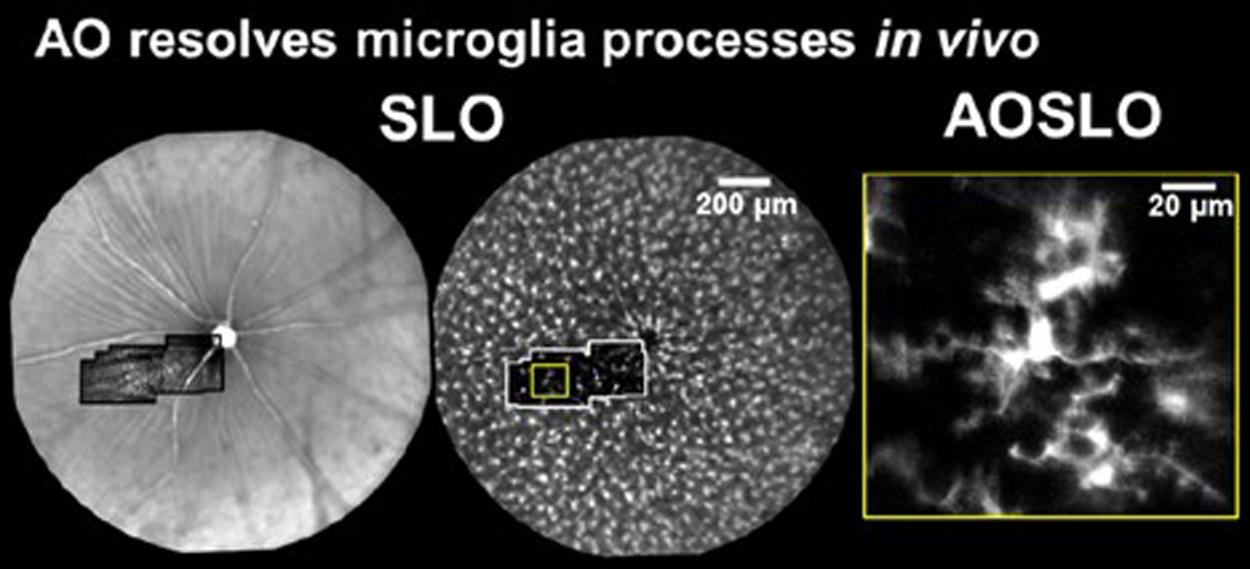
Microglial processes are highly motile while cell somas were relatively very stable over the course of hours. Process motility was quantified using strategies similar to that used for quantifying blood cell speed. Process velocity moves at a snail’s pace at the range of 0-6.4 µm/s (Schallek et al. ARVO e-abstract 2017), similar to the range reported in the brain.
Microglial cells can be tracked from milliseconds-to months. Cell processes are highly motile during minutes to hours, cell somas can also be observed to migrate during weeks to months.
![]()
Related Publications
Aby Joseph, Derek Power, Jesse Schallek (2021) Imaging the dynamics of individual processes of microglia in the living retina in vivo. Biomed Opt Express. 2021 Sep 10;12(10):6157-6183. https://doi.org/10.1364/BOE.426157
Imaging immune cell movement as a biomarker for retinal inflammation
The microenvironment of the eye provides a unique space for immune cells to interact with the vascular system, the neural parenchyma and the overall ocular health of the subject. We want to explore whether the motility of immune cells can be considered as a biomarker for the ocular microenvironment to proceed or initiate the inflammatory response.
After an inflammatory cue is encountered, there are changes in retinal blood vessels that allows them to become selectively more adhesive in response to signaling from the resident retinal immune cells. This enables the immune cells in blood circulation to slow down at the blood vessel endothelium, that eventually allows them to enter the retina.
When we track the movement of these single immune cells that are slowing down at the vascular walls of the retina, we observe a variety of behaviors ranging from loosely adhered cells that roll along the vein wall, to tightly adhered crawling cells. Very rarely, we observe cells that are actually migrating against the direction of blood flow. (Dholakia et al. ARVO abstract 2022)
We would also like to know if these immune cells move at a constant speed along the vein wall, or are there regions along the vein where they slow down more than others. We have found preliminary evidence of such microdomains of high and low adhesion. This has promising implications for the next step in the immune extravasation cascade - where the systemic immune cells exit blood circulation and enter the retinal tissue. (Dholakia et al. ARVO abstract 2023)
With our AOSLO, we can image the kinetics of these cells when they are freely flowing in blood circulation, after they bind to the vein wall and even after they enter the retina. The circulating cells move at incredibly fast speeds ranging in millimeters/seconds. The slow moving rolling and crawling leukocytes at the vein endothelium that move at micron/seconds speeds. Then we had the extravasated leukocytes that were moving at a glacial pace of microns/minutes. These behaviors are vastly different- in fact, the speed of leukocytes in circulation is five orders of magnitude higher than their speed after they extravasate into the retina. (Dholakia et al. 2022 Fall Vision Meeting abstract). These findings also indicate that imaging the anatomy and speed of immune cells may serve as a new cellular biomarker of inflammation.