

Tissue-on-a-Chip Tech Could Speed Cures from Clinical Trials
When he was a kid, Ben Miller used to read a lot of science fiction.
“I don't anymore,” he said, “because I'm living it.”
Miller is part of a group of researchers developing small, bioengineered devices with core elements measured in micrometers to mimic human cells and organs. This tissue-on-a-chip technology promises to revolutionize medical research trials as we know them today.
The hope is to improve on, and one day even replace, mouse experiments that have been the standard of scientific inquiry for the last 100 years. When perfected, the technology could limit animal testing as well as speed clinical trials and avoid the potential catastrophe of a human trial that causes unforeseen harm to participants.
The worldwide pursuit of tissue-on-a-chip devices that are reliable and robust enough to replicate human biology has been accelerated in the U.S. through federal mandates, along with grants to a handful of select academic research powerhouses including Duke, Harvard, Johns Hopkins, and the University of Rochester.
URochester actually started late. But in this race to redefine how diseases are studied and how emerging treatments are tested, a small group of Rochester researchers and students now has an inside track.
Looking at how the work has developed here, it's not surprising that URochester is a leader in this field; in several crucial ways, it could only have happened here.
Big Change Coming—Quickly
Tissue chips are bioengineered, microfluidic devices embedded with human cells in a way that keeps them alive for days or even weeks. They mimic physical, biological, and even mechanical environments of human tissue and organs so that scientists can observe cell interactions and responses when studying disease or testing potential cures. This could largely replace current testing for disease modeling, drug discovery, toxicology screening, and even personalized medicine.
University of Rochester researchers Hani Awad, James McGrath, and Miller are leading federally funded tissue-on-a-chip research projects on the River and Health Sciences campuses. Combined, these researchers have nearly a century of fruitful research to their names, but all three describe tissue-on-a-chip as the biggest development in their careers.
They see the coming change profoundly affecting academia, industry, and medicine. And soon.
Even the rare successful trial is lengthy. “In the best of all worlds, a drug makes it to market after 13 to 15 years of development,” said Miller, a Dean's Professor of Dermatology at Rochester with joint appointments in Biomedical Engineering, Biochemistry and Biophysics, Optics, and Materials Science.
The lack of human-relevant data slows the process.
“Animal studies do not sufficiently predict a drug's effects,” explained Awad, the Donald and Mary Clark Distinguished Professor in Orthopaedics and a professor of Biomedical Engineering.
Differences in reactions between mice and humans are a problem, along with the cost, labor, and time to care for living animal subjects. And inbred research mice lack the genetic diversity of humans.
Frequently, drugs have been shown to be safe and effective in animal trials but not in human subjects.
Pushed by those failures and pulled by the promise of emerging technologies, the FDA issued a 2025 mandate to reduce and then largely replace animal testing for drug development by 2029. It earmarked $150 million to develop, test, and standardize human-based research models such as tissue-on-a-chip, organoids, AI, and computer modeling.
The NIH has now established a new Office of Research Innovation, Validation, and Application (ORIVA) to reaffirm its commitment to advancing scientifically valid alternatives to animal testing, and it has a “Complement Animal Research in Experimentation” program to facilitate New Approach Methodologies (NAM) development.
URochester is in the thick of all this. Early work included research by Ben Frisch, PhD, assistant professor of Pathology and Laboratory Medicine, to build tissue chips for studying acute myeloid leukemia, and Lisa DeLouise, PhD, associate professor in Dermatology and in Biomedical Engineering, to identify drugs to protect salivary glands from radiation damage during cancer treatment.
In 2020, URochester became one of 10 NIH-funded research centers around the nation for testing tissue chips' potential to simulate clinical trials. It received $4 million to develop a tissue chip to test therapies for tendon injury.
In 2024, NIH awarded URochester a $7.5 million grant to establish the Translational Center for Barrier Microphysiological Systems (TraCe-bMPS) in partnership with Duke University to build FDA-qualified drug development tools. Powered by URochester technologies and industry and academic partnerships, the project is using tissue chips on Rochester-built platforms to study human models in the muscle-immune system, neuroinflammatory injury, joint-lining remodeling, immune cell trafficking, and the blood-retinal barrier.
In the same year, URochester earned a three-year contract valued at up to $7.1 million from the Biomedical Advanced Research and Development Authority (BARDA), within the Department of Health and Human Services, to model respiratory disease effects on the brain and test drugs to prevent and treat symptoms. This will open a new way to study the effects on the brain from long COVID and viruses such as influenza.
Progress is already taking the work outside the lab and attracting industry.
Made in Rochester
McGrath, Miller, and Awad have founded Rochester-based chip-manufacturing companies that are producing more than 5,000 tissue chips and more than 50,000 photosensors per year, supplying 12 URochester labs and nine others around the world. These labs have at least 35 chip projects underway, and the number keeps growing.
URochester has developed some game-changing advantages with its approach.
McGrath's nanomembrane, for one, is the thinnest available, which helps the cells behave more naturally. Thicker materials used by other researchers give a cloudy aspect to cells seen through a microscope, but “Jim's nanomembrane is optically transparent … as if it is not there,” Awad said. And with such a thin layer, there's no interference from a membrane between cells. “We can embed Ben's photonic sensors in the chip to measure what these cells are releasing in real time.”
Many chips on the market are made of a stretchy, elastomeric material that can leach or absorb substances used in testing. URochester has developed chips that use silicon nanomembranes and photonic sensors in a hard plastic shell, minimizing the leaching problem.
The chips are also modular, so a multitude of users can customize them for different areas of research. The chips give researchers insight into human cell behavior that they never had before. They have channels to simulate blood flow across tissue or to introduce pathogens such as viruses to trigger an immune response in the cells, or a biologic agent that can affect an inflammatory response, or a drug in development to test its benefit or toxicity. The chips can be engineered with posts to have changeable structures in order to alter the amount of mechanical force in the microenvironment. This “stretches” the cells to simulate the breathing motions of a lung, the shear forces of blood flow, or the mechanical stress on a tendon.
“Modularity in the design is absolutely a competitive advantage,” McGrath said. “Our tissue chips are very different than anything else out there. It's very difficult for others to reproduce what we do. And this is all Rochester homegrown technology.”
The pharmaceutical industry has noticed. AbbVie, Pfizer, Bausch + Lomb, and ApoGlyx have a vested interest in streamlining the drug development process and are working with URochester; AbbVie and Bausch + Lomb also provide funding. Non-pharma industry partnerships include manufacturers and system developers such as SiMPore, ALine, Phlotonics, SiObex, and AIM Photonics. Academic partnerships are surging as well. Duke University and Rochester Institute of Technology are part of the consortium in the TraCe project; other academic centers involved with URochester include the University of Michigan, University of Oregon, Georgia Tech, and Indiana University.
The sheer volume of research at multiple sites means standardization of processes is essential. A team led by Joan Adamo, PhD, director of the Office of Regulatory Support in the Clinical and Translational Science Institute, is developing quality systems as a blueprint for researchers to follow. More than 60 standard operating procedures (SOPs) have been published to date.
SOPs for tissue chip research are vital to the integrity of findings and another form of leadership in an emerging industry.
“This is such a new approach, we are having to invent as we go,” Adamo said. “We don't have 30 to 40 years of standard practice for this type of research.”
Human-equivalent chips, Awad said, “would create safer and more effective drugs faster. … Predicting failed drug candidates early in the development process would save pharmaceutical companies tons of money and potentially shorten the development process from 13 or 15 years to maybe 7 to 10.”
But there is a long road ahead. The chips need to use a few hundred cells to accurately replicate the human body's response, all while keeping cells alive long enough to get meaningful results. A way to analyze and catalog the voluminous data from testing must be perfected. And as with all manufacturing, scaling up is a challenge.
URochester researchers are racing to find solutions, and in many critical aspects they are at the head of the pack. That in itself is remarkable, considering that the roots of this technology began elsewhere. In Rochester, it took several fortunate twists of fate and even a failed experiment to leap ahead. And it might not have happened at all if not for a student who connected some dots and changed several people's lives.
The Johnnys-Come-Lately
In 2018, graduate student John Cognetti started working in Miller's lab. But he also worked in McGrath's lab. And something clicked.
He described it this way:
“I said, ‘I like what Jim's doing with microphysiological systems, but I like Ben's sensors too; I would like to put them together because it seems like most of the models out there don't really do much in the way of sensing.’”
Everyone agreed it was a good idea.
“So then I spent the next five or six years trying to get it to work,” he said.
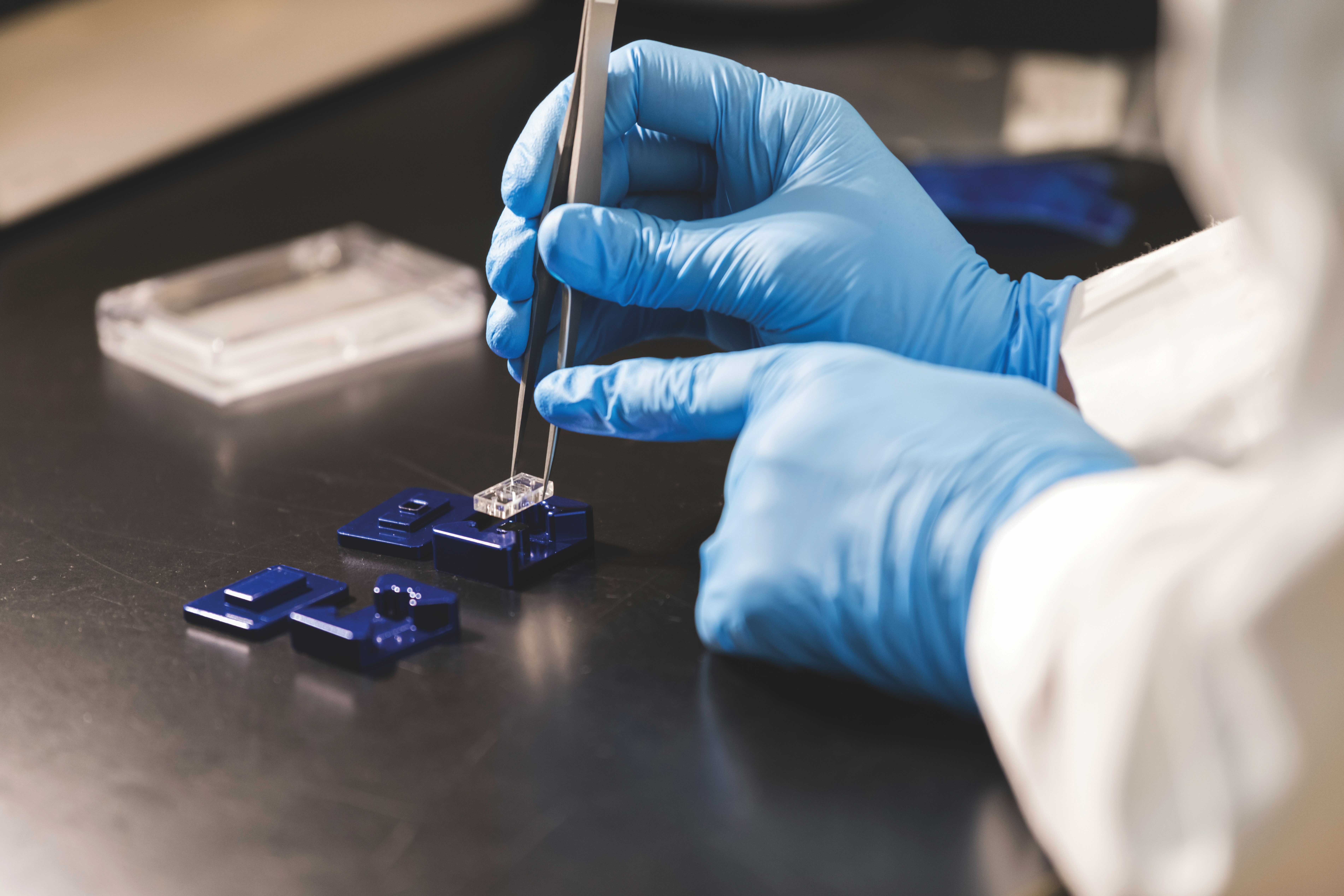
Cognetti had to address many engineering details in order to get technologies to work together. He had to adapt what he knew about microfluidics to work with Miller's sensors, which use laser light to measure cell activity. He also spent a lot of time figuring out how to affix McGrath's nanomembrane to the chip, eventually turning to a precision craft cutter.
In other words, URochester was a relative newcomer. Yet the pieces were in place.
“Tissue-on-a-chip is an area my lab has been interested in forever, in some ways going back to when I was a postdoc,” Miller said.
The collaboration between labs was the catalyst.
A Louis Pasteur Moment
Chance favors the prepared mind, Pasteur said. Case in point, the breakthrough nanomembranes came about by accident. And the photonic sensors were developed for a different purpose altogether.
The nanomembranes were born out of a lab project gone wrong: A post-doc, Chris Striemer, was trying to make a photonic conductor out of a silicon layer.
“He kept making it thinner and thinner and thinner to help visualize silicon crystals,” said McGrath, “but when he baked it at a thousand degrees (Celsius), in addition to the silicon crystals, he saw nanopores. The pores ruined it for his application, but Chris was smart enough to realize the material could have utility.”
So he brought it to McGrath's lab.
“We designed some experiments to show that the membranes were really exceptional at filtering molecules,” said McGrath.
So exceptional that the results were published in Nature.
“That changed our lives and we went all in. I pivoted my career to focus on applications of this new material.”
It took nearly a decade of work to enhance the raw material, understand it, and make it stable enough for lab use. Blood-brain barrier researchers began reaching out to McGrath to try it.
“So I knew there was value in the material for those researchers, and we built it as a tissue chip for them. It's a relationship that continues to this day.”
Likewise, the photonic sensors were not originally built for tissue chips.
Miller, who has been at URochester for 30 years, had been developing sensors that proved useful as a diagnostic tool during COVID. They're now used in tissue chips to enable real-time measurement of cell activity.
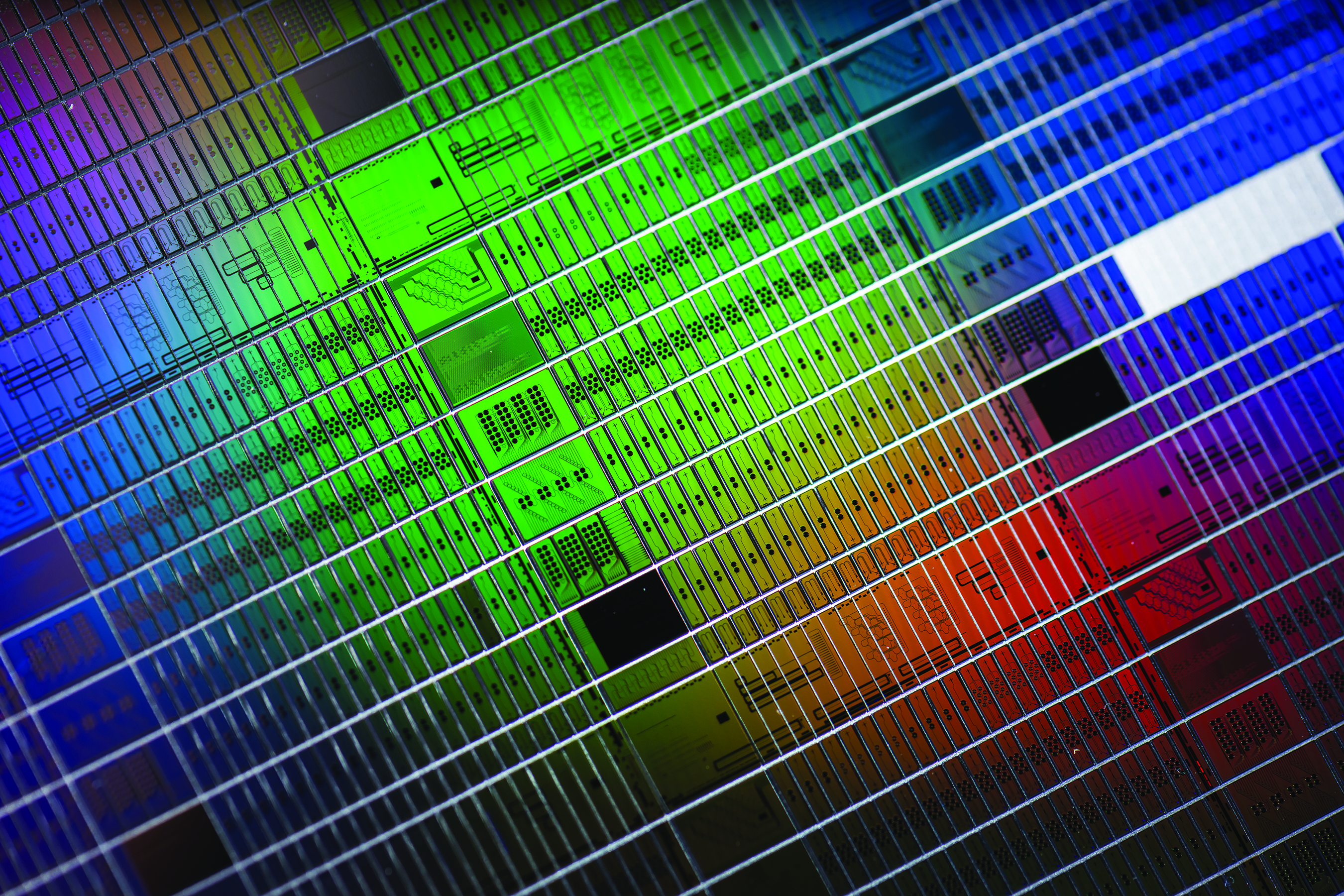
Each sensor has eight to 18 different individual microring resonators that use tiny beams of light to detect a specific cell activity. When a cell starts secreting something in response to a drug or toxin, the device gives a reading that notes the change.
Manufactured in Albany, the photosensors arrive as 11,000 individual units fabricated on a single disc the size of a vinyl LP record.
In the lab, the team then builds the tissue chips with components tailored to the needs of each experiment.
“We use the nanomembranes to culture the cells and circulating media in the device to keep them alive,” Miller said.
The photosensors measure and record cell activity in real time, unlike other chips that require disassembly and an end to the experiment to determine how cells reacted.
The researchers seed the chips with a variety of cell types.
A brain chip, for example, uses different types of donated cells from different parts of a person's brain.
“You've got admittedly a very simple model,” Miller said, “but a model of that person now on a chip.”
That model brings a clear benefit.
“We have been able to see differences from donor to donor in drug response for an anti-inflammatory. And that starts to be super exciting because the way drug development is done now, you don't see any of that until the phase one clinical trial.
“Doing that on chips, you already have an idea of how it's going to work before it's actually used in part of a human being.”
This has implications for theranostics, a personalized approach to medicine that combines diagnostic imaging and targeted therapy specific to an individual's diagnosis.
“We could take that a step further,” Miller said. “You can have the theranostic procedure but then also test a panel of different drugs for a person to find the most effective drug for their condition and genetic makeup.”
For the promise of personalized medicine, tissue-chip technology would enable research projects to solicit donor cells from a true cross-section of the population to achieve better outcomes.
Males, females, pregnant people, all age ranges, and all races could be represented in any clinical trial by donating a small tissue sample, giving researchers a picture of how a potential drug would work for a variety of patients.
And potentially, each person could contribute donor cells for a personalized study to see which cancer drug could be most effective for them.
Chips Changing Research Now
Tissue-on-a-chip “is big because it can be applied to any problem,” said Frisch of Pathology and Laboratory Medicine.
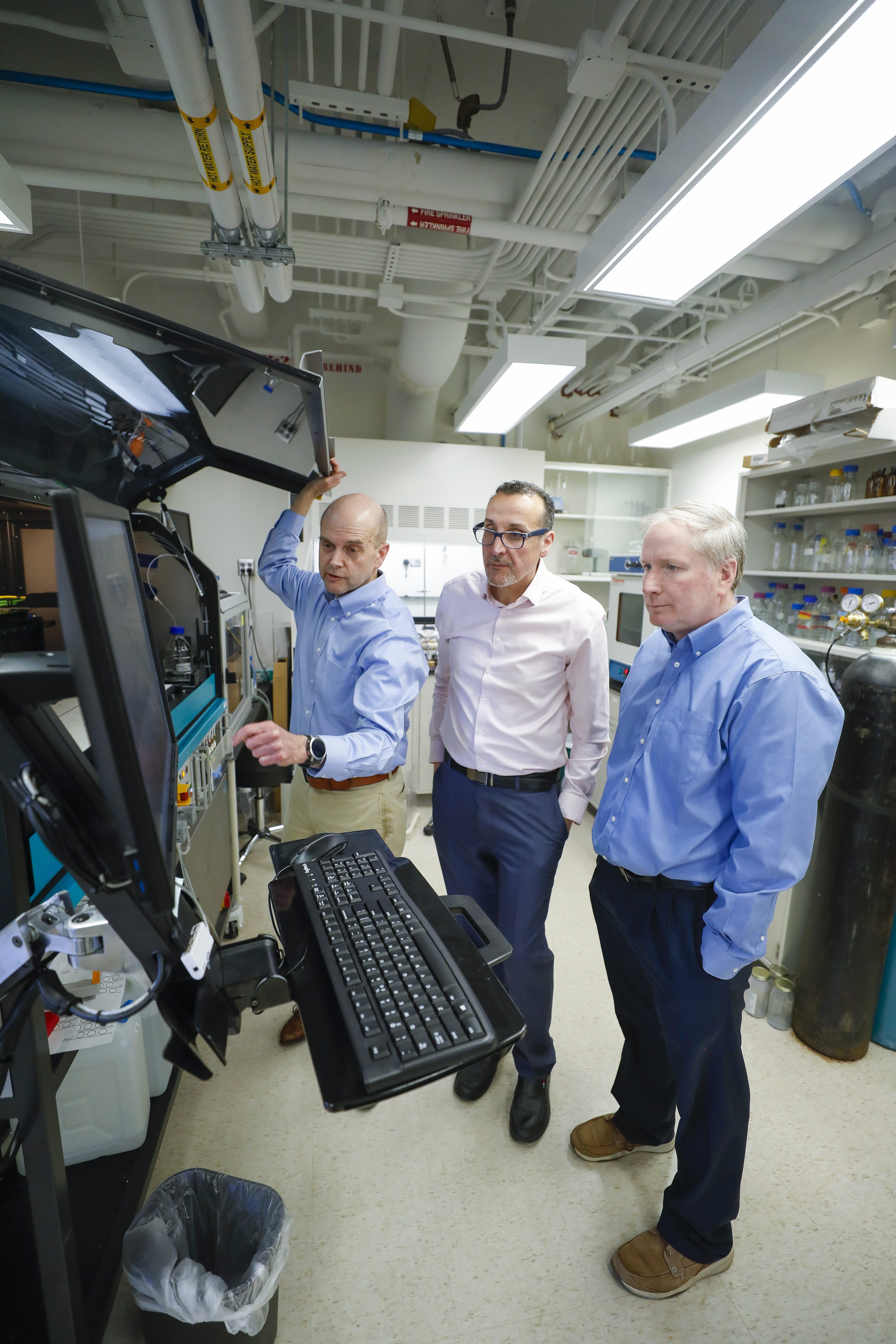
Frisch is using tissue chips to model leukemia, and this is especially useful because cancer medications can be very toxic to human tissues.
Using chips has important advantages over current single-cell studies in a dish, Frisch said.
“Cells on a dish are not getting any of the interaction or communication that they normally get from other cells in their environment. And that can fundamentally alter the way they behave and the way they respond to therapies.”
In a dish, he explained, it's easy to figure out what can kill cells and control their growth.
“But they often respond completely differently when they have that support in an environment that they're used to growing in. With tissue chips, we can provide a lot more of that interaction … and they will respond more like what they would in their native environment in the human body.”
The tissue chips he uses can simulate blood flow through vessels and the exchange of nutrients such as oxygen and carbon dioxide across a cell layer, out to the tissue and back.
He also has built an extracellular matrix made of fibroid gel and other cell types critical for normal blood production and cancer cells.
“All of that, on one chip,” he said.
The hope is to save time, money, and patients' efforts by testing different possible therapies on tissue chips first, then choosing the most effective to take into clinical trials.
But it's not just an advantage in cancer research.
Awad sees the same thing with tendon-on-a-chip technology that URochester is developing:
“We shouldn't be looking at that one cell particularly. We should be looking at the microenvironment within which it lives.”
In the tendon chip, a collagen hydrogel simulates an injured tendon's microenvironment, which includes tendon cells and macrophages.
The gel keeps cells in their “scar” microenvironment.
A top compartment has the nanomembrane containing the single-layer endothelial cells that line blood vessels, allowing researchers to simulate circulating blood cells.
The nanomembranes are the building block of Rochester's chips, and they are a game-changer.
“It is interest from others that affirms that our platform has unique value,” McGrath said. “No one is approaching tissue-on-a-chip this way.”
But just like the importance of needing a microenvironment for better testing, there will be no “one chip fits all” solution.
“When the NIH began to think about replacing animal models, they weren't just thinking about tissue chips,” Awad said. “It's really a multitude of complementary technologies.”
In 2017, DeLouise of Dermatology and Biomedical Engineering received a $3.8 million NIH grant to support her invention of microbubble array technology.
Circular 3-D chambers are an ideal environment in which to culture and study groups of cells, and the cupped shape of her device facilitates cell interaction.
She's using it to study preventive treatments for salivary gland radiation damage sustained by head and neck cancer patients.
It's another example of expanding the breadth of what is discoverable, a key focus of URochester's vision.
The TraCe project, for example, shows the value of URochester's emphasis on linking different types of tissue chips.
“Our modular platform design grew out of the fact that we were building different chips to meet different needs,” McGrath said. “I realized that we couldn't support collaborations with a single chip geometry, so we created a platform where the parts could be redesigned and ‘snapped' together by non-engineering laboratories.”
What that means for the future is where things begin to look more like science fiction.
Your Own Personal Chip?
As the research is developed, Awad said, “We can think about potentially creating your own tissue chip made out of your own cells.”
He imagines taking a skin biopsy, then using fibroblast cells from the connective tissue and inducing them into stem cells.
On your chip, those cells could grow into whatever is needed.
“We can create your immune cells, we can create your cartilage, bone, whatever, right? So the technology is here, we have protocols, and we've been perfecting them. It ultimately comes down to scale. Can we scale this up to enable these kinds of experiments?”
Scaling these tiny tissue chips requires thinking big.
“We need colleagues to become partners and drive the adoption of these tissue chips,” Awad said. “We think this can be accomplished by establishing an institute for microphysiological systems at the University of Rochester to do work similar to institutes at powerhouses like the Whitehead Institute.”
The risk is losing ground.
“We certainly get a lot of interest from other universities and researchers wanting to take aspects of our technology and integrate it into theirs,” Miller said.
“The really key thing is continuing to build these models and validate them against whole animal models and human disease and health. That's something that needs to happen here.”
Already, the pace of URochester's acceleration into more advanced tissue-chip systems has been possible only because of the multiple grants it has received.
That, and a relentless sharing of ideas.
“I think it's a particular strength of the University of Rochester: We're a culture of collaboration,” Miller said. “My lab and Hani's are at the medical center, and Jim's lab is just across the street. We have all these different people we can talk to all the time. For 30 years, I've been saying this to people: We recognize that we're a relatively small place and for us to have an impact, we know that we have to work together. So we're motivated to do that ... We all want to work together.”
Connect with us!





Author
