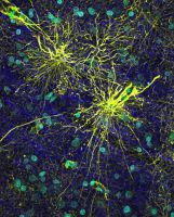
Astrocytes Help Separate Man from Mouse

A type of brain cell that was long overlooked by researchers embodies one of very few ways in which the human brain differs fundamentally from that of a mouse or rat, according to researchers who published their findings as the cover story in the March 11 issue of the Journal of Neuroscience.
Scientists at the University of Rochester Medical Center found that human astrocytes, cells that were long thought simply to support flashier brain cells known as neurons that send electrical signals, are bigger, faster, and much more complex than those in mice and rats.
“There aren’t many differences known between the rodent brain and the human brain, but we are finding striking differences in the astrocytes. Our astrocytes signal faster, and they’re bigger and more complex. This has big implications for how our brains process information,” said first author Nancy Ann Oberheim, Ph.D., a medical student who recently completed her doctoral thesis on astrocytes.
The study is one of the most extensive examinations yet of the astrocyte. Oberheim and co-authors discovered a previously unknown form of the cell, a varicose projection astrocyte, in the human brain but not in the rodent brain. The team also found that the most abundant type of astrocyte, protoplasmic astrocytes, are approximately 2.6 times larger than their rodent counterparts, and that the human cells have about 10 times as many “processes,” or structures designed to connect to other cells.
“We have not really been able to understand why the human brain is so much more capable than that of any other animal,” said neuroscientist Maiken Nedergaard, M.D., D.M.Sc., who led the study. “Some people have thought that it’s simply that a bigger brain is a better brain, but an elephant’s brain is bigger than a person’s, for example, but it’s not nearly as powerful. So that’s not the answer.
“It may be that humans have a much higher brain capacity in large part because our astrocytes are more sophisticated and have more complex processing power,” added Nedergaard, who spoke last week at a Gordon Research Conference on glial biology. “Studies in rodents show that non-neuronal cells are part of information processing, and our study suggests that astrocytes are part of the higher cognitive functioning that defines who we are as humans.”
Astrocytes had long been considered passive support cells, a means to hold the rest of the brain cells together, like glue. Medical students might spend a few minutes pondering the astrocyte before moving on to their flashy counterparts, the neurons that fire the electrical signals crucial to pretty much everything we do. It’s the electrical activity of neurons that constitutes what most scientists have considered to be brain activity, and it’s the neurons that are the target of every currently available drug aimed at brain cells. If astrocytes were important, scientists thought, it was most likely because they help create a healthy environment for the neurons.
It turns out that astrocytes, which are 10 times as plentiful as neurons, had been pushed to the boundaries of neuroscience because of a gap in the tools used to study the brain. Scientists measure signaling among brain cells mainly by looking at electrical activity. But astrocytes don’t fire in the same way as neurons, and so conventional techniques don’t record their activity. So when scientists “listened” with conventional techniques, they witnessed no activity.
Rather than realizing their tools were incomplete, scientists assumed that astrocytes were silent.
So Nedergaard devised a new way to “listen” for astrocyte activity, developing a sophisticated laser system to look at their activity by measuring the amount of calcium inside the cells. Her team has discovered what might be called the secret lives of astrocytes and has made a series of startling discoveries. Astrocytes use calcium to send signals to the neurons, and the neurons respond; neurons and astrocytes talk back and forth, indicating that astrocytes are full partners in the basic working of the brain; and astrocytes are central to conditions like stroke, Alzheimer’s, epilepsy, and spinal cord injury.
“Dogma is slow to change, and one of the dogmas of neuroscience is that astrocytes are support cells that don’t do much themselves,” said Oberheim. “The view is slow to change, but scientists are coming around. Astrocytes are now acknowledged as active participants in brain function and sensory processing.”
The brain’s two signaling systems – one composed of neurons, and one of astrocytes – complement each other, Nedergaard said. Neurons send signals extremely quickly over long distances – the hand touches a hot stove, for instance, and the brain detects the danger and moves the hand away, instantly. Astrocytes, in contrast, send slower signals whose function is still being worked out by scientists.
“The brain contains two communication networks using different languages,” said Nedergaard, director of the Division of Glial Disease and Therapeutics of the Center for Translational Neuromedicine. “You have a highly sophisticated electrical network embodied in the neurons, which send signals instantaneously. And then you have a much slower network composed of astrocytes whose signals are 10,000 times slower but which might be able to process the information in a more sophisticated manner and retrieve memories.
“There is no other tissue in the body that mixes up two different types of cells so completely as how astrocytes and neurons are interspersed throughout the brain,” Nedergaard added. “Both comprise extensive signaling networks. Where those networks interface and how they interact makes the brain so interesting.”
To do the study, the team studied human brain tissue taken from 30 people who had had surgery, mostly to treat epilepsy or brain tumors. They compared the astrocytes in human brains to those in mice and rats. In addition to the findings above, the team noted additional differences:
- Astrocytes in people signal five times as fast as those in mice and rats.
- Human astrocytes are organized into more complex units known as domains than are rodent astrocytes. A typical rodent domain includes tens of thousands of neuronal synapses, while the team found that a human domain might include up to 2 million synapses. These domains are highly organized groupings of cells that appear to be precisely situated, almost like atoms in a crystal. This organization is likely important for information processing, said Nedergaard, who notes that brain injury is associated with a loss of astrocytic domain organization and a decrease in cognitive function.
- In people, cells known as fibrous astrocytes, which are mainly for structural support, are on average more than twice as large as their counterparts in mice and rats. Also, people have another type of cell known as interlaminar astrocytes, which are not present in rodents.
- Another difference concerns the end feet of protoplasmic astrocytes, which wrap around blood vessels throughout the brain and are thought to play a role in the brain’s blood flow. In humans, these end feet cover the walls of blood vessels much more completely than they do in mice and rats, possibly playing a much more important role in keeping agents in the blood from entering the brain and in regulating blood flow.
The authors include Steven Goldman, M.D., Ph.D., professor and chair of Neurology; Webster Pilcher, M.D., professor and chair of Neurosurgery; Jeffrey Wyatt, D.V.M., professor and chair of Comparative Medicine; and research assistant professors Takahiro Takano, Ph.D., Xiaoning Han, Ph.D., Wei He, Ph.D., and Fushun Wang, Ph.D.
Other authors include technical associate Qiwu Xu; Jane Lin, Ph.D., of New York Medical College; and Jeffrey Ojemann, M.D., and Bruce Ransom, M.D., Ph.D., of the University of Washington, where Oberheim completed part of her doctoral thesis under Nedergaard’s supervision.
The work was funded by the G. Harold and Leila Y. Mathers Charitable Foundation and by the National Institute of Neurological Disorders and Stroke.

