ECG Placement
Videos
View videos and instructions for EKG lead placement.
EKG Quality Improvement Project
Maher Abadeer, MD, 3rd Year Fellow, Pediatric Cardiology and Jan Schriefer, M.B.A., M.S.N., Dr.P.H., Director of the Quality and Patient Safety Program
Situation
There is significant room for improvement when obtaining ECGs. The improvements can lead to more accurate diagnostic information which may result in more timely and accurate diagnosis.
Background
ECGs are important modalities with which to diagnose structural heart abnormalities and arrhythmias in children. They can also be important indicators of cardiac muscle injury or inflammation. The percentage of ECGs needing improved technique in Golisano Children’s Hospital varies by unit. Some units perform a large volume of ECG’s yet have room for improvement.
Assessment
We have found that in inpatient units in the children’s hospital, the ECG completion error rate is between 18-36%. This is compared to a lower rate of 3.5% in the cardiology clinic. Up to a third of these errors are limb lead reversals, which can be easily corrected with proper lead placement. The majority are due to poor ECG tracing quality and artifact which may be due to patient movement and/or other environmental and external stimuli or mechanical support. We have also discovered that poor quality of ECGs may not always be documented in the final read by the pediatric cardiologist. Finally, the current administrative nursing ECG protocol needs updating, including the correct EMRN to use.
Recommendation
Our hope is to improve ECG tracing quality with two methods:
- By improving detection by attending cardiologists using a review presentation and laminated cards to serve as a resource, outlining different types of lead reversals and the proper documentation coding for each.
- Improving nursing and technician ability to prevent errors and troubleshoot with the use of available photographs and diagrams near each ECG machine, “Just in Time” videos demonstrating lead placement for each type of patient body habitus, raising awareness and education on minimizing artifact, and updating/correcting the current ECG nursing protocol.
Instructions
Prior to Lead Placement
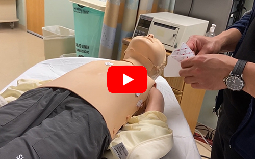
- Please input the "EMRN," by typing "E###..." (Use a capital "E")
- For neonates, please use patient’s LEGAL name (e.g. “boy” or “girl”)
- Ensure lead/cable connection and lead/box connections are correct
- Ensure skin is dry so leads stick (e.g., try alcohol swabs)
- May need to cut lead stickers as instructed so they don’t touch
- Please allow > 2 min to settle patient to minimize ECG artifact
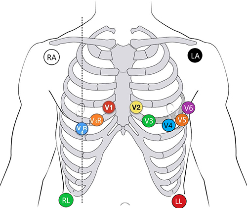
Lead Placement
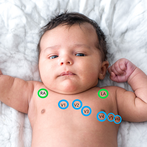
- V1 – 4th intercostal space to the right of the sternum
- V2 – 4th intercostal space to the left of the sternum
- V3 – Midway between V2 and V4 (left side)
- V4 – 5th intercostal space at the midclavicular line (nipple line) left side
- V5 – 5th intercostal space, anterior axillary line
- V6 – 5th intercostal space, mid-axillary line
- V7 – 5th intercostal space, just beyond V6
- V3R – Midway between V1 and V4R on right side, (if V3R leads available)
- V4R – 5th intercostal space at the midclavicular line (nipple line) right side (if V4R leads available)
- Right Arm (RA) and Left Arm (LA) limb leads placed on wrist or above elbow
- Right Leg (RL) and Left Leg (LL) limb leads placed between knee and ankle
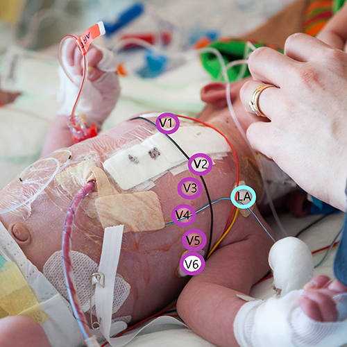
Note: On babies and toddlers who can’t keep their legs and arms still:
- Put leg leads on lower abdomen near right & left iliac crest.
- Put arm leads 1-2 inches below the right & left clavicle.
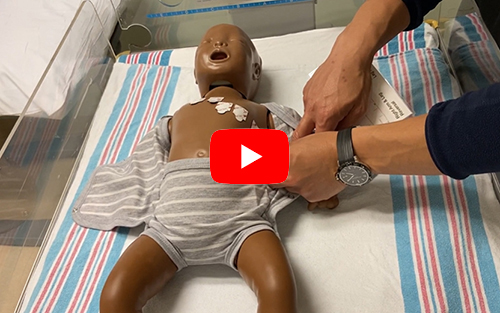
ECG Lead Placement
Infant
Video: Demonstration of proper ECG lead placement on an infant mannikin and troubleshooting steps.
To view larger images click an image or link.
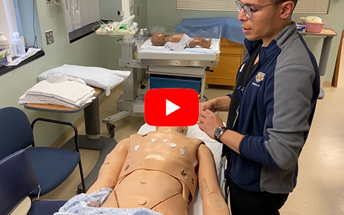
Young Child/Toddler
Video: Demonstration of proper ECG lead placement on a young child mannikin and troubleshooting steps.
Older Child/Adult
Lead stickers can be placed below the clavicle rather than the shoulder to prevent muscle twitch artifact.
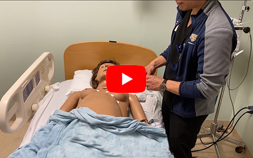
Female
Video: Demonstration of proper ECG lead placement on a female mannikin and troubleshooting steps.
Lead stickers can be placed below the clavicle rather than the shoulder to prevent muscle twitch artifact.
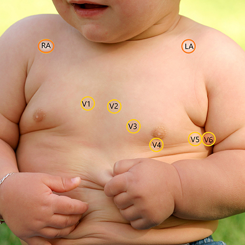
Young Child with Obesity

Older Child / Adult

Female - Frontal

Female - Side