
Investigating Developmental Origins, Mechanisms and Therapies for Retinopathy of Prematurity Using an In vivo Mouse Model of Oxygen-Induced Retinopathy
The Developing Retina and Oxygen
Retinopathy of prematurity (ROP) is a bi-phasic condition of abnormal retinal vascularization in premature infants exposed to supplemental oxygen, characterized by dysregulation of vascular endothelial growth factor (VEGF). Although the etiology of ROP is multifactorial, the most significant risk factor is oxygen.
Our eyes provide a unique window through which retinal blood vessels can be directly visualized. Premature infants are being born at earlier gestational ages (as early as 22 weeks gestation) due to new medical technologies and clinical practice strategies, including antenatal steroids for lung maturity, post-natal surfactant replacement in immature lungs, gentler modes of ventilation methods, improved nutrition, and prevention of infection. However, premature birth exposes the developing retinal blood vessels to harsh external stimuli and a relatively hyperoxic ex utero environment compared to the physiologically hypoxemic in utero conditions. Hyperoxia disrupts retinal blood vessel development, leading in subsequent weeks to pathologic neovascularization, bleeding and retinal detachment.
Serial eye exams in the neonatal intensive care unit (NICU) help identify at-risk infants for earlier treatment with retinal laser photocoagulation and/or intravitreal anti-VEGF injections. Current ROP therapies are limited in efficacy due to structural/functional adverse effects or systemic toxicities, therefore ROP remains one of the leading causes of childhood blindness worldwide.
In vivo Retinal Imaging in ROP
The Mezu-Ndubuisi lab studies the effects of oxygen on the retina using newborn mice, who at full term birth are at the same developmental stage as premature babies born at 24 to 26-week gestation. Knowing that the retina can be visualized through an optically clear media, we discovered a way of studying ROP in mice without extracting the retina, leading to the development of an in vivo mouse model of OIR.
In this model, we perform live imaging of the dilated eyes of anesthetized mice after oxygen exposure to directly visualize their retinal blood vessels revealing unique retinal vascular abnormalities (arterial tortuosity, venous dilation, and capillary avascularity) more distinctly at different stages of development. We developed customized objective methods for quantitative analysis of these retinal vascular features, www.quantbv.com. Our studies led to the novel use of in-vivo phosphorescence lifetime imaging to measure retinal vascular oxygen tension, fluorescein angiography (FA) to depict retinal vascular abnormalities, and spectral-domain optical coherence tomography (SD-OCT) to correlate retinal thickness. We have simultaneously correlated in vivo vascular and structural changes in mice with OIR to functional abnormalities that could lead to neuronal dysfunction using electroretinogram (ERG), as well to an abnormal long-term retinal histologic phenotype of prolonged cellular apoptosis, persistent micro and macroglial activation, and formation of ectopic synapses.
The Mezu-Ndubuisi lab is currently exploring molecular mechanisms of oxygen-induced retinopathy, and the cross-talk between angiogenesis and inflammation in disorders of developing organs of premature babies in the retina, brain, lungs, and kidneys. We are also investigating innovative anti- and pro-angiogenic therapies that could be safer, non-toxic therapies for ROP, and potentially applicable to other diseases of prematurity, in order to improve long-term neuro-developmental outcomes of fragile premature infants.
Schematic of in vivo and Histologic Phenotypes in ROP and OIR
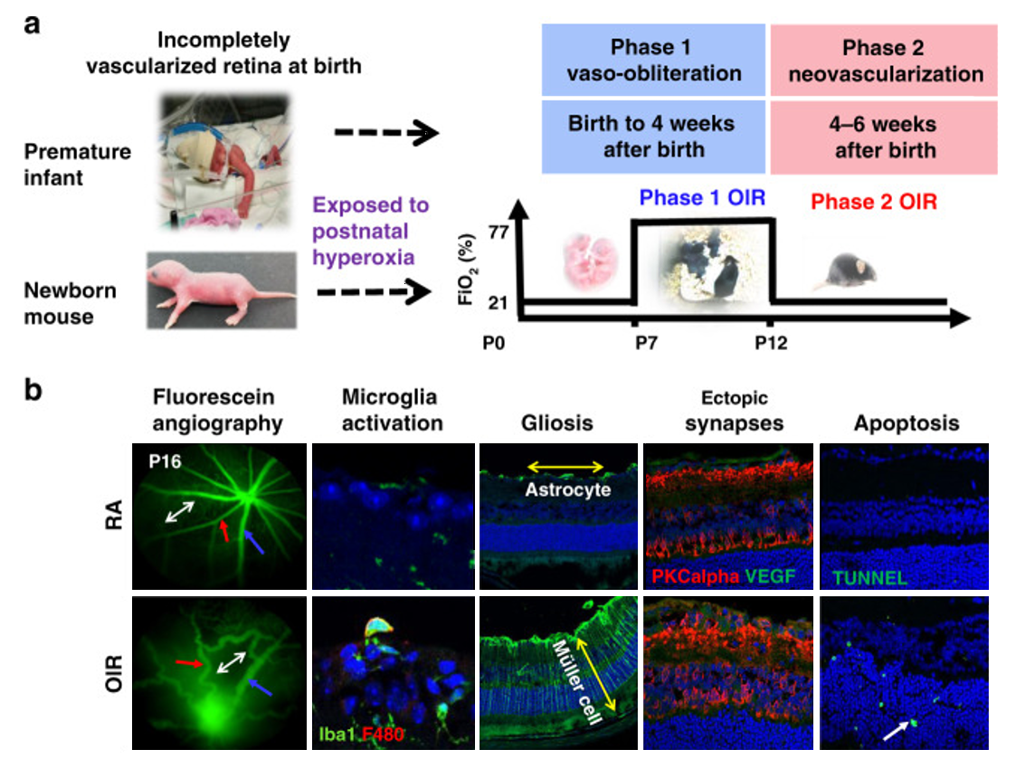
a. The newborn mouse and the premature infant both undergo postnatal retinal development. Postnatal hyperoxia triggers Phase 1 ROP (vaso-obliteration). Hypoxia leads to Phase 2 ROP (neovascularization).
b. In vivo imaging of P16 using fluorescein angiography. Blue arrows: veins; red: arteries; white double arrow: capillary network. Histology shows persistent ectopic synapses, neuronal apoptosis, and gliosis in RA and OIR mice. The yellow double arrow shows astrocytes, while the yellow double arrow shows Müller cells. The short white arrow represents TUNEL+ cells. Iba1 stains microglia. PSDS stains photoreceptor postsynaptic terminals, PKCalpha stains bipolar cells. ROP retinopathy of prematurity, TUNEL terminal deoxynucleotidyl transferase dUTP nick-end labeling, P postnatal day age.
Published in: Mezu-Ndubuisi OJ, Song YS, Macke E, Johnson H**, Nwaba G**, Ikeda A, Sheibani N. Retinopathy of prematurity shows alterations in Vegfa164 isoform expression. Pediatr Res. 2021 Jul 20. doi: 10.1038/s41390-021-01646-9. PMID: 34285351.
McPherson Eye Research Institute's Retina Research Foundation Edwin and Dorothy Gamewell Professor
Associate Professor of Pediatrics and Ophthalmology, University of Rochester, Rochester New York
Lab Home >

Olachi Mezu-Ndubuisi, M.D., O.D.
Principal Investigator
- Retinal Vascular Recovery revealed by retinal imaging following neonatal hypoxia ischemia in mice: is there a role for tyrosine kinase receptor modulation?; Brain research. 2022 Sep 15.
- Role of the Endothelium in Neonatal Diseases.; Newborn (Clarksville, Md.); Vol 1(1), pp. 44-57. 2022 Mar 31.
- Community Preventive Health Education Intervention for Pediatric Iron-Deficiency Anemia in Rural Southeast Nigeria.; Annals of global health; Vol 88(1), pp. 105. 2022 Jan 21.
- Retinopathy of prematurity shows alterations in Vegfa164 isoform expression.; Pediatric research; Vol 91(7), pp. 1677-1685. 2021 Jul 20.
- Unmasking Systemic Racism and Unconscious Bias in Medical Workplaces: A Call to Servant Leadership.; Journal of the American Heart Association; Vol 10(7), pp. e018845. 2021 Mar 29.
Affiliations
- Department of Pediatrics, University of Rochester
- Department of Ophthalmology, University of Rochester
- Flaum Eye Institute, University of Rochester
- McPherson Eye Research Institute, Wisconsin
Lab News
Mezu-Ndubuisi Lab at the 2025 APHA Conference
Congratulations to the Mezu-Ndubuisi Lab global health students for four research abstracts selected for presentations (one oral and three posters) at the American Public Health Association (APHA) conference in Washington DC, November 2-4, 2025.
View All Lab News
Contact Us
Olachi Mezu-Ndubuisi, M.D., O.D.
Principal Investigator, Mezu-Ndubuisi Lab
Flaum Eye Institute, Department of Ophthalmology
University of Rochester Medical Center
575 Elmwood Avenue
Rm 1.3011, Box 659
Rochester, NY 14642
Lab Shipping Address
Mezu-Ndubuisi Lab
University of Rochester Medical Center
Department of Ophthalmology
Flaum Eye Institute (Room 1-3022B)
University of Rochester,
Rochester, NY 14642
(585) 275-0562 Flaum Eye Institute Inquiries
Lab Home >