MRI Equipment and Resources
Magnet
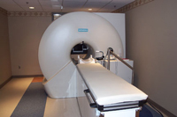
The heart of the URCABIN is a Siemens 3T MRI MAGNETOM PrismaFit whole-body scanner (software version VE11c) recently upgraded (fall 2017) with maximum gradient amplitude of 80 mT/m and a slew rate 200T/m/s for each gradient direction. There are a 20-channel and a 64-channel phased array head coils (capable of parallel imaging using SENSE/GRAPPA) which are available for brain studies. In addition there are a whole array of specialized coils for body, extremity and other scans.
Pulse sequences installed on the PrismaFit system allow capability for many types of research applications, including functional MRI (EPI-BOLD), conventional structural MRI (T1 and T2 weighted imaging), diffusion-weighted scans including diffusion tensor imaging (DTI and other advanced techniques for imaging white matter tracts in the brain), Arterial Spin Labeling (ASL) perfusion, Susceptibility Weighted Imaging (SWI) and Quantitative Susceptibility Mapping (QSM), single- and multi-voxel spectroscopy, among others. Fast imaging techniques such as simultaneous multislice (SMS) is available for DTI, EPI-BOLD, and other advanced sequences.
The center has a research agreement with Siemens Medical System to provide researchers with access to pulse sequences beyond standard protocols and access to on-going Investigator-requested Prototype for Research (IPR), or Consumer-to-Producer (C2P) software packages. Customized RF coils may be designed and fabricated with support from Siemens by local experts.
Mobile Brain/Body Imaging
The Mobile Brain/Body Imaging system, or MoBI, combines virtual reality, brain monitoring, and Hollywood-inspired motion capture technology, enabling researchers to study the movement difficulties that often accompany neurological disorders and why our brains sometimes struggle while multitasking.
Using the same technology that is employed by movie studios to produce CGI special effects, study participants wear a black body suite that is fitted with reflective markers. Participants are then asked to walk on a treadmill or manipulate objects at a table in a room fitted out with 16 high speed cameras that record the position of the markers with millimeter precision. This data is mapped to a computer generated 3D model that tracks movement.
Pulse Sequence Programming
All of the standard pulse sequences used for clinical protocols are available as part of the Siemens 3T system. In addition, custom pulse sequences can be programed for specialized applications via a research agreement with Siemens and by local researchers. Several faculty and graduate students have attended the Siemens pulse sequence programming (IDEA) course offered at their US training facility in North Carolina.
Custom Coils
Custom RF coils can be designed and fabricated for specialized projects when the standard human head coils, and other coils at URCABIN are not appropriate. On-going research and development has produced coils with new concepts and designs that include a liquid nitrogen cooled phased array coil and a phased array coil for monkey brain imaging. Additional information can be found on RF Coil Development site.
Integrated Stimulus Equipment
The scanner control room is outfitted with a Virtually Silent workstation running Windows and Ubuntu Linux that is interfaced to the scanner (for synchronization) and to the LCD video display in the scan room (BOLDscreen, Cambridge Research Ltd) and audio equipment (SereneSound Digital, Resonance Technology Inc.) to present stimuli to participants as they are being scanned.
The presentation computer also samples button-press responses (PI Engineering X-Keys keyboard system and Cambridge Research fORP) and can record subject voice responses to collect data for those protocols that use such responses.
The BOLDscreen LCD display is calibrated and capable of displaying video with a refresh rate up to 120Hz at a resolution of 1920x1080 without any buffering, ideal for very fast stimuli or 3D applications.
The SereneSound audio system (Resonance Technology Inc.) features and active noise cancellation patient microphone, a flexible headset that conforms to the shape of the head coil, 30dB gradient noise attenuation, 40Hzto 40kHz frequency response, high fidelity speakers for the MRI suite, and a multifunctional remote control.
Standard software packages for stimulus display and response recording (e.g., PsyScope, Presentation, E-Prime, Direct RT, Psychophysics Toolbox, and MATLAB/Octave) are preloaded or available on request. Stimulus presentation on the computers can be synchronized with the MRI scanner.
MRI compatible optical goggles are supplied (MediGlasses, Cambridge Research Ltd) to provide vision correction to the subjects for visual stimulus.
Also, a physiological measurement system, also synchronizes with the MRI scanner (BIOPAC Systems, Inc.). The recording measurements currently available are ECG; Respiration; CO2 gas analysis, Pulse and SPO2. This system comes equipped with AcqKnowledge® software which allows researchers to identify the frequency of the gradient noise of the MRI scan and then remove it from the signal. The gradient artifact can be removed in real-time and during post-acquisition processing.
Mock Magnet
 The URCABIN mock magnet can be used to train research subjects how to perform an experiment without using any time on the Prisma magnet. The mock magnet has setup mirrors that resemble the Prisma as closely as possible, with an audio-visual presentation system for aural and visual experiments, and a surround sound system that reproduces the noises the actual MRI scanner makes during a scan.
Data Storage and Management
Data obtained from the scanner is automatically sent to URCABIN data storage servers and an archive copy is made at that point. Users are responsible for accessing, quality control and long-term storage of their own data.
The University has various storage methods available such as High-Availability Enterprise Storage (SMD-NAS), Cloud Storage (Box) as well as High Performance Storage Systems at CIRC which URCABIN can integrate with.
Every PI is responsible for creating and maintaining a proper Data Management Plan (https://dmptool.org/) associated with their grants and IRB protocols. In order to create accounts and receive data, you will need to send a copy of this DMP with the IRB approval or an IRB waiver. If you intend to share this data outside the University of Rochester, a Data Usage Agreement with each institution has to be in place and shared with the URCABIN well prior to accessing the data.
If you have questions regarding data storage and data use, please contact support@rcbi.rochester.edu