

Neuroscience and Innovation: Redefining Stroke Care & Recovery
Every minute after a stroke, nearly two million brain cells die. But increasingly, researchers and clinicians are demonstrating that stroke care is not only about restoring blood flow in the first critical hours; it is also about understanding how the brain repairs, reorganizes, and recovers in the weeks and months that follow.
At URochester Medicine, that work spans the entire continuum of stroke care, from mobile emergency treatment and regional telemedicine networks to research exploring brain plasticity, rehabilitation, clot biology, and the roles of sleep and circadian rhythms in recovery. These efforts are helping shape a more personalized and neuroscience-driven approach to stroke treatment and recovery.
Building a Regional System of Stroke Care
URochester Medicine’s clinical stroke program is built around a regional system of care that begins before patients reach the hospital and extends through emergency treatment, intensive care, rehabilitation, and long-term recovery. At the center of that system is the Comprehensive Stroke Center at Strong Memorial Hospital, which includes the Neuromedicine ICU and Acute Brain Injury Rehabilitation Unit and brings together specialists in stroke neurology, neurosurgery, neurocritical care, rehabilitation medicine, and imaging.
One of the program’s most visible innovations is the Mobile Stroke Unit (MSU). Introduced in 2018, the specially equipped ambulance functions as an emergency department on wheels, carrying a CT scanner, point-of-care laboratory testing, telemedicine capability, and clot-busting medications directly to patients in the field. Earlier this year, URochester Medicine expanded the program with a second MSU, extending the reach of rapid stroke treatment across the region.
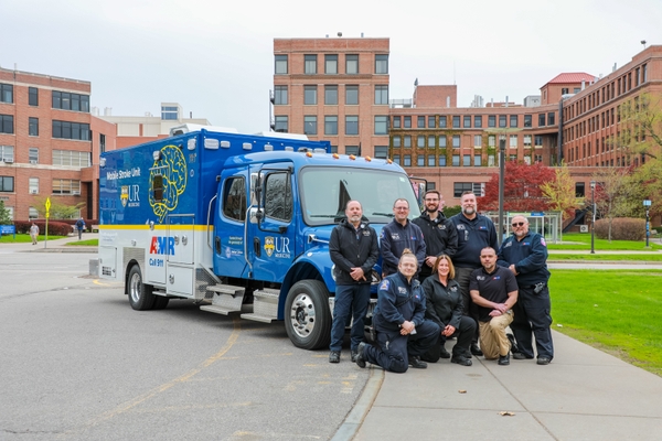
From left: Frank Post, Nate Coons, Joshua Lesperance, Jason Burgett, Nathan Young, Lena Brockmann, Zachary Callahan, and Justin Connolly are members of URochester Medicine/AMR Mobile Stroke Unit team.
“The Mobile Stroke Unit allows us to deliver specialized stroke care at the earliest possible moment, before a patient even reaches the hospital,” said neurosurgeon Tarun Bhalla, MD, PhD, director of the MSU program. “The second unit builds on years of field experience, expands access to rapid diagnosis and treatment, when every minute can mean the difference between recovery and disability.”
The Telestroke program extends that reach even further. Led by neurologist Adam Kelly, MD, the service provides 24/7 remote stroke and neurological consultations for 16 hospitals across upstate New York and now fields more than 4,000 calls annually. Through video consultations and rapid review of imaging and clinical data, stroke specialists help determine which patients require advanced interventions—such as thrombectomy, ICU care, or complex vascular procedures—and which can remain at their local hospital.
The model strengthens stroke expertise while preserving highly specialized resources for the patients who need them most. It also creates a powerful platform for research by connecting investigators to a large, diverse patient population and by accelerating the translation of new discoveries into clinical care.
“What makes our stroke program unique is that we cover the entire spectrum—from basic neuroscience to innovative recovery strategies, clinical trials, and a high-volume stroke care network that reaches patients across the region,” said neurologist Curt Benesch, MD, medical director of the Comprehensive Stroke Center. “We’re not just treating the stroke in front of us; we’re constantly working on how to prevent the next one, expand acute treatment options, and make sure that evidence-based care reaches every bedside.”
Relearning Movement After a Stroke
Stroke recovery does not end when a patient leaves the hospital. Increasingly, researchers at URochester Medicine are showing that the brain retains a remarkable ability to adapt, relearn, and reorganize after injury—a property known as plasticity— and that understanding those mechanisms may transform rehabilitation.
By understanding how the brain adapts after injury, scientists are developingtherapies that are more personalized.
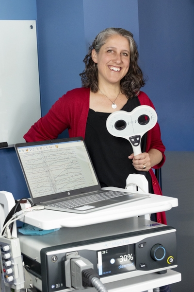
In the Movement and Plasticity Lab, stroke neurologist Ania Busza, MD, PhD, studies how the brain relearns movement after stroke and how rehabilitation can better harness that recovery process. Her work focuses on motor system plasticity, the way practice and experience reshape surviving brain circuits after injury. “Recovery after stroke isn’t just about getting stronger,” Busza said. “It’s about helping the brain learn how to use the remaining circuits it still has.”
For decades, rehabilitation often followed a relatively simple principle: more therapy leads to better outcomes. But researchers now recognize that stroke patients develop very different motor control problems depending on which neural pathways are damaged. Some struggle with delayed movement, others with fatigue or involuntary activation of opposing muscles.
“Two people may both have weakness after a stroke, but the underlying control problem may be completely different,” Busza said. “If we understand those differences, we can start designing rehabilitation that is much more personalized and targeted.”
Her lab combines neuroscience, imaging, biomechanics, engineering, and rehabilitation science to better understand those differences. In one project, patients use faint electrical signals from weakened muscles to control a simple video game through surface electromyography (EMG). Even patients unable to lift their arms can participate. The system helps researchers track how motor impairments evolve during the first months after stroke and identify early warning signs of long-term disability.
Researchers are also studying whether interventions such as vagal nerve stimulation, transcranial magnetic stimulation, and other neuromodulation approaches can safely amplify the brain’s natural plasticity and improve recovery.
The broader goal is to understand not only whether patients recover, but also how recovery occurs within the brain itself. Modern neuroscience has changed how researchers think about that process. Rather than the brain simply “rewiring” around damage, scientists now believe recovery depends on whether alternative, partially redundant circuits can be strengthened and refined through practice.
“Practice after stroke is really a form of learning,” Busza said. “The brain is constantly adapting based on experience, and rehabilitation is trying to shape that adaptation in a productive way.”
Restoring Vision After a Stroke
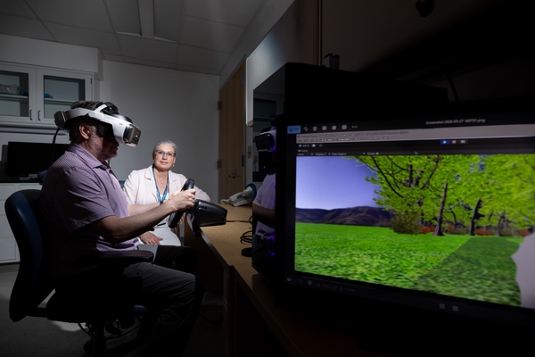
Krystel Huxlin, PhD, is studying another often-overlooked consequence of stroke: vision loss. Her research focuses on patients whose strokes damage the visual cortex in the occipital lobe, leaving them partially blind. Historically, many of these patients were told little could be done. “There has been a longstanding belief that vision loss after stroke is permanent and untreatable,” Huxlin said. “Our work has shown that it is not true.”
Huxlin’s Visual Perceptual Restoration Laboratory has demonstrated that intensive visual training can help patients recover some visual abilities in damaged portions of the visual field. The approach works much like physical therapy for movement recovery, in which repeated, carefully structured practice trains surviving brain circuits to process visual information more effectively.
“We’re essentially retraining the visual system,” Huxlin said. “The damaged brain still has some capacity to change, but you have to engage it in the right way.”
The work has implications beyond vision rehabilitation. Huxlin says it reflects a major shift in neuroscience itself. A few decades ago, the idea that the adult brain could meaningfully reorganize after injury remained controversial. Today, plasticity is recognized as a lifelong feature of the nervous system.
Her lab now uses advanced imaging, eye tracking, state-of-the-art virtual reality headsets, and brain stimulation to better understand the mechanisms behind visual recovery and make rehabilitation more accessible to patients at home.
Together, the work of Busza, Huxlin, and their collaborators reflects a larger transformation in stroke research. By understanding how the brain adapts after injury, scientists are developing therapies that are more personalized, more targeted, and increasingly grounded in the biology of recovery itself.
What Stroke Clots Can Reveal
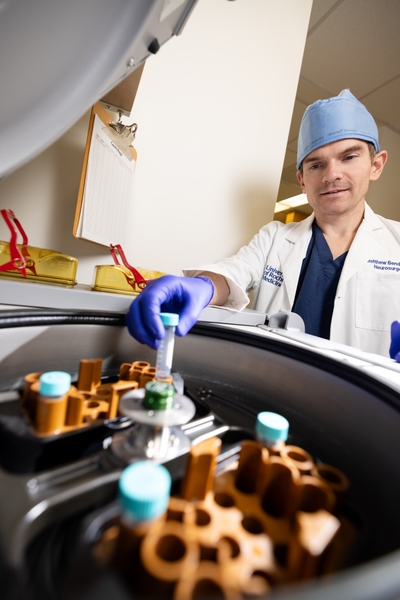
To neurosurgeon Matthew T. Bender, MD, the clot removed during a stroke procedure is more than medical waste—it is a source of critical information about why strokes happen and how patients respond to treatment.
Bender’s research focuses on the molecular composition of stroke clots, particularly in patients with large-vessel occlusion strokes treated with thrombectomy. By analyzing proteins and inflammatory markers within these clots, his team hopes to better understand why some strokes are harder to treat and why outcomes vary dramatically between patients.
“Some stroke researchers find the clots themselves to be bland or uninteresting,” Bender said. “We feel the opposite very much.”
His work has identified proteins linked to more difficult clot removal, worse recovery, and complications such as hemorrhage. The long-term goal is to develop blood-based biomarkers that could help physicians tailor stroke treatment to individual patients.
Conducting that work at a high-volume stroke center is critical. URochester Medicine performs more than 200 thrombectomies each year, giving researchers access to blood and clot samples that would otherwise be discarded.
“Those biospecimens are invaluable,” he said. “They allow us to study how clots form in real human stroke patients and develop better ways to break them down.”
The Role of Sleep, Timing, and the Brain’s 'Cleaning System'
Stroke has traditionally been viewed as a problem of blocked blood flow, but emerging research suggests it may also be a disorder of timing.
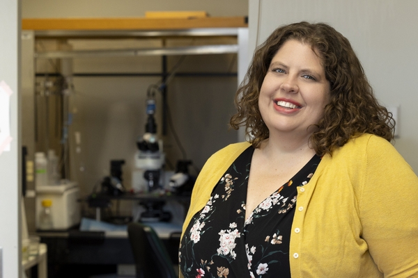
Neuroscientist Lauren Hablitz, PhD, studies how sleep, circadian rhythms, and the brain’s glymphatic system—a network that clears waste from the brain during sleep— influence stroke recovery. The glymphatic system, first discovered in the lab of URochester Medicine neuroscientist Maiken Nedergaard, MD, DMSc, appears to become impaired after stroke, potentially allowing inflammatory molecules and cellular waste to accumulate.
“Our central question became: if timing is broken after stroke, can we improve recovery by reinforcing the biological clock?” Hablitz said.
Using mouse models, researchers tested interventions designed to strengthen circadian rhythms, including timed feeding and experimental clock-targeting drugs. Both approaches improved glymphatic function, reduced inflammation, and improved recovery after stroke.
Rather than viewing recovery solely as an inflammation problem, Hablitz believes researchers may also need to consider the brain’s ability to clear harmful signals and metabolic waste. “If the system responsible for clearing signaling molecules is not working properly,” she said, “everything builds up.”
The findings suggest that relatively simple interventions— such as timed eating, light exposure, or therapies that improve sleep and circadian health—could one day become part of stroke rehabilitation strategies.
Author
