Abdominal Imaging
Make Appointments & Get Care
Overview
When you need answers about abdominal pain, digestive symptoms, or other concerns affecting the organs and blood vessels in your abdomen, accurate imaging is an important first step. The abdominal imaging specialists at University of Rochester Medicine use advanced technology to help diagnose conditions, guide treatment decisions, and monitor your health over time.
Our Approach
URochester Medicine offers full range of abdominal imaging services, including CT, MRI, ultrasound, fluoroscopy, and image-guided procedures. Our experienced radiologists work closely with your care team to provide timely, accurate results so you can move forward with confidence.
Computed Tomography (CT)
Computed tomography (CT) uses X-rays to create detailed cross-sectional images of the abdomen and pelvis. CT scans help diagnose injuries, infections, tumors, kidney stones, digestive conditions, and many other abdominal disorders.
Our imaging centers use state-of-the-art multi-detector CT scanners and advanced image reconstruction software to produce high-quality images while completing exams as quickly as possible.
We perform a full range of CT studies, from routine abdominal and pelvic imaging to specialized exams such as cardiac calcium scoring. Our radiologists also work closely with Nuclear Medicine specialists to interpret PET-CT examinations when needed.
Magnetic Resonance Imaging (MRI)
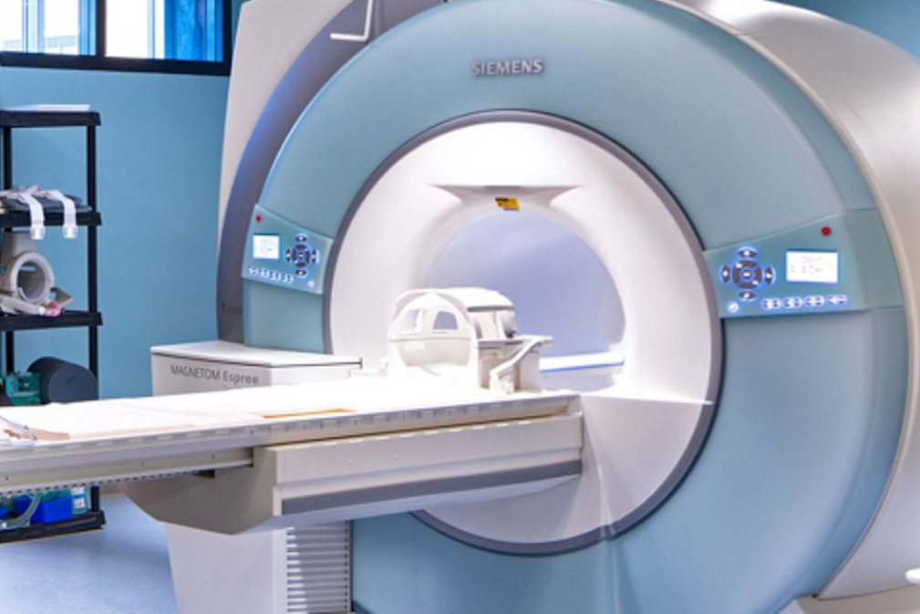
Magnetic resonance imaging (MRI) uses powerful magnets and radio waves to create detailed images of organs, soft tissues, and blood vessels without using radiation. MRI can help diagnose a wide range of abdominal conditions and provide information that may not be visible on other imaging tests.
Our inpatient and outpatient imaging locations are equipped with advanced MRI technology for routine and complex examinations. Specialists perform advanced MRI techniques, including MR angiography, functional MRI, and specialized imaging sequences that help deliver accurate diagnoses.
Our imaging team also supports ongoing MRI research, with expertise in cardiac MRI and emerging imaging technologies that continue to advance patient care.
Ultrasound
Our abdominal imaging specialists use advanced ultrasound technology to evaluate organs, blood vessels, and blood flow throughout the abdomen. Ultrasound uses sound waves to create detailed images without radiation, making it a safe and effective way to diagnose and monitor many abdominal conditions.
We have six sonography rooms equipped with real-time, color flow Doppler, and other state-of-the-art equipment. Our experts perform vascular, body, neurosonography, and ultrasound-guided interventional procedures, including:
Abdominal Aortic Ultrasound
The standard imaging test for the abdominal aorta is an ultrasound, primarily used to find aneurysms.
Abdominal Doppler Ultrasound
An abdominal Doppler ultrasound evaluates blood flow in the veins or arteries in the abdomen.
Mesenteric Doppler Ultrasound
A mesenteric doppler ultrasound is a specialized test that examines blood flow of the veins or arteries in the stomach and intestines.
Gastrointestinal and Genitourinary Imaging
The Gastrointestinal and Genitourinary (GI/GU) Imaging subdivision provides specialized imaging to diagnose conditions affecting the digestive and urinary systems. Our radiologists work closely with referring providers to deliver accurate,timely results that help guide treatment decisions.
Our team performs specialized imaging studies, including:
- Single- and double-contrast gastrointestinal examinations
- Enteroclysis for detailed evaluation of the small intestine
- Three-phase pharyngograms to assess swallowing disorders
- Excretory urography
- Cystography
- Voiding cystourethrography (VCUG)
- Retrograde urethrography
- Hysterosalpingography (HSG)
Our faculty radiologists and residents also interpret abdominal and pelvic digital radiographs, providing expert imaging evaluation for a wide range of gastrointestinal and genitourinary conditions.
Not sure what to expect from an imaging exam?
Learn how to prepare, what happens during your visit, and how imaging helps your care.
What Sets Us Apart?
Choosing where to receive abdominal imaging matters. At University of Rochester Medicine, our patients benefit from the expertise of an academic health system where fellowship-trained radiologists work closely with specialists across many disciplines to diagnose and manage conditions affecting the abdomen and pelvis.
Our team offers:
- Fellowship-trained abdominal imaging radiologists with advanced expertise in interpreting complex imaging studies
- Comprehensive abdominal imaging using CT, MRI, ultrasound, and image-guided techniques
- Close collaboration with gastroenterology, hepatology, transplant surgery, oncology, urology, gynecology, emergency medicine, and primary care providers
- Expertise in diagnosing conditions involving the liver, pancreas, kidneys, gastrointestinal tract, spleen, and other abdominal and pelvic organs
- Image-guided biopsies and minimally invasive procedures performed by Interventional Radiology specialists when appropriate
- Access to advanced imaging, specialized procedures, and coordinated specialty care—all within one integrated health system
By combining advanced technology with coordinated, patient-centered care, we help deliver accurate diagnoses and personalized treatment plans.