Dense Breasts
Make Appointments & Get Care
What Are Dense Breasts?
Dense breasts are common and can be found in almost half of women over the age of 40. Dense breast tissue is composed of milk glands, milk ducts, supportive tissue (dense breast tissue) and fatty tissue, which can make it harder to detect cancer on a mammogram.
UR Medicine Breast Imaging offers:
The Tyrer-Cuzick Risk Assessment Calculator, a nationally accepted standard based on science, helps to measure women’s breast cancer risk factors based on family history, and helps women to better understand their breast cancer risk factors. It allows health professionals to estimate a woman’s risk of developing invasive breast cancer over the next five years and up to age 90.
*This tool can’t accurately estimate breast cancer risk for women:
- Carrying a breast-cancer-producing mutation in BRCA1 or BRCA2
- With a previous history of invasive or in-situ breast cancer
- Lobular cancer in- situ (LCIS)
- Ductal cancer in-situ (DCIS)
- In certain other subgroups
What Causes Dense Breast Tissue?
It’s not clear why some women have a lot of dense breast tissue and others do not; however, you are more likely to have dense breasts if you are in your 40s or 50s, are premenopausal or take hormone therapy for menopause. For most women, breasts become less dense with age.
How Does Dense Breast Tissue Look on a Mammogram?
When viewed on a mammogram, dense breast tissue appears as a solid white area, which makes it difficult to see through. Breast tissue that is not dense appears dark and transparent. Women with dense breasts have an increased risk of getting breast cancer. The denser the breast, the greater the risk.
Why is Additional Imaging So Important for Women with Dense Breasts?
Dense breasts can make it harder to detect cancer on a screening mammogram. Although screening mammograms can detect cancers, experts often recommend additional screening methods to more accurately make a diagnosis.
UR Medicine's Treatments for Dense Breasts
3D Mammography/Tomosynthesis
Tomosynthesis mammography, also known as 3D mammography, is a new method of screening for breast cancer. The test involves taking several X-rays of each breast from various angles to create a detailed 3D image.
Tomosynthesis finds invasive cancers at a 40% higher rate than regular mammograms. It can also pinpoint hard to find cancers that may otherwise be unnoticed, particularly in areas of dense tissue.
Ultrasound
A breast ultrasound is a non-invasive imaging test that uses high-frequency sound waves to create detailed images of the structures within the breast. It is used to help check for breast abnormalities, especially in telling the difference between solid masses and fluid-filled cysts.
Contrast Enhanced Mammogram
A contrast enhanced mammogram (CEM) uses a special contrast dye to highlight abnormal areas in breast tissue. The process is similar to a standard mammogram, with the addition of an injection of the contrast to create clearer, more detailed images. This type of mammogram is especially helpful for detecting breast cancer in people with dense breast tissue or when other imaging results are unclear. It is available at Strong Memorial Hospital and our Red Creek breast imaging location in Henrietta.
MRI
A breast MRI (magnetic resonance imaging) creates detailed images of the breast tissues using large magnets and a computer. Unlike X-rays, MRI does not use radiation. Providers often use breast MRIs to check known breast cancer. Breast MRIs are often used with mammograms and ultrasounds to help find and diagnose breast cancer and other breast health issues. This service is offered at our offices at East River Road in Rochester, Breast Imaging Center in Canandaigua and St. James Hospital Medical Office Building in Hornell.
What Sets Us Apart?
The UR Medicine Breast Imaging Center is designated a Breast Imaging Center of Excellence by the American College of Radiology’s (ACR) Commission on Quality and Safety and the Commission on Breast Imaging for a three-year term. The ACR Breast Imaging Center of Excellence designation signifies that UR Medicine Breast Imaging provides these services to the greater Rochester area and surrounding communities at the highest standard of radiology profession.
Our expertise is backed by research. As part of an academic medical center, our providers are continuously working to advance the detection and assessment of breast disease. We invest in cutting-edge technology.
We collaborate with breast specialists at UR Medicine’s Wilmot Cancer Institute to ensure that all necessary treatments and support is available.
Locations
View All LocationsWe serve you in the Rochester metropolitan area and surrounding region.
View All Locations7 locations
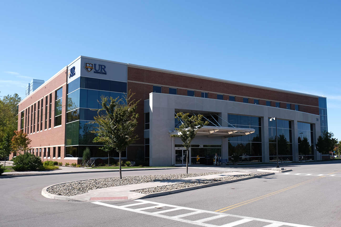
200 East River Road
Rochester, NY 14623
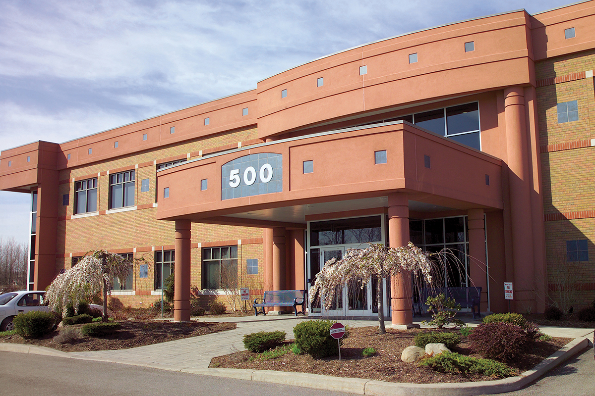
Calkins Corporate Park (Red Creek)
500 Red Creek Drive, Suite 130
Rochester, NY 14623
Lakeside Professional Park
229B Parrish Street, Suite 103
Canandaigua, NY 14424
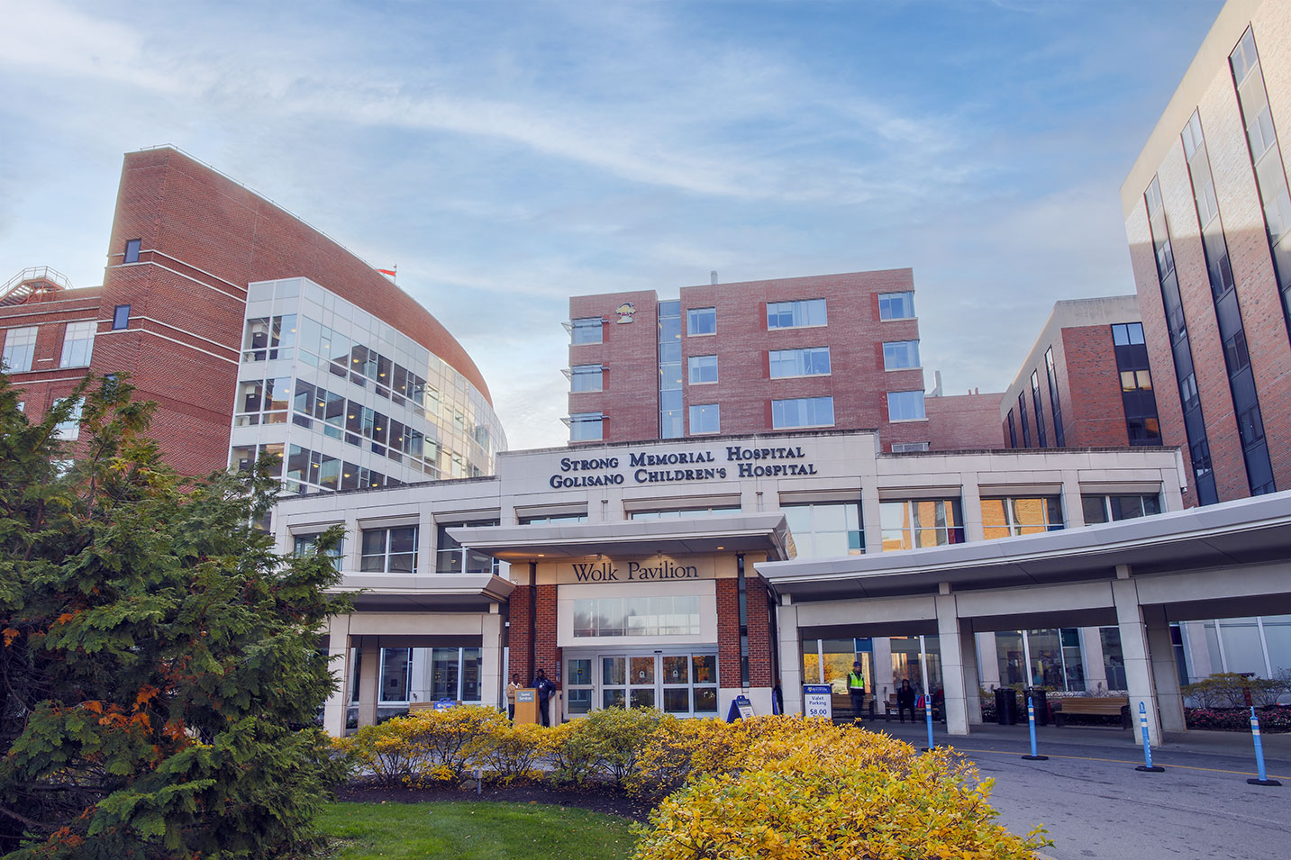
Strong Memorial Hospital
601 Elmwood Avenue
Rochester, NY 14642
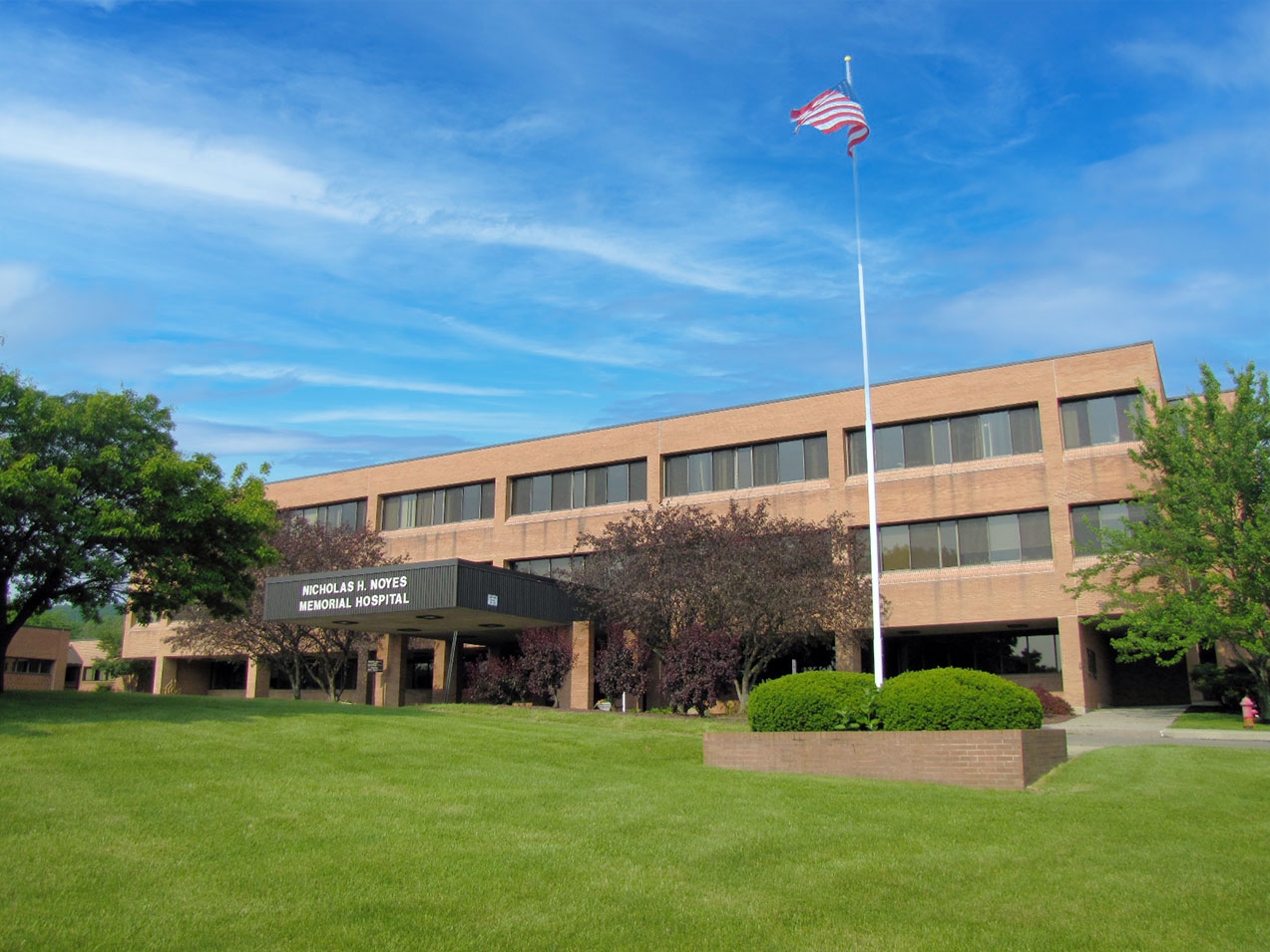
E. Michael Saunder Medical Imaging at Noyes Memorial Hospital
111 Cara Barton Street
Dansville, NY 14437
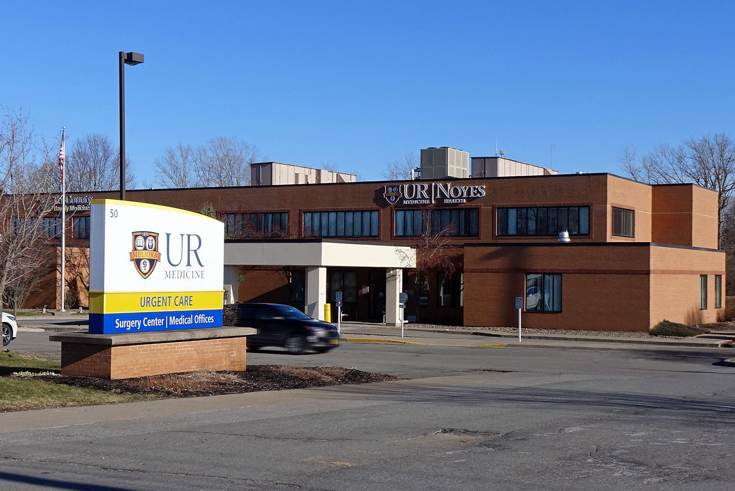
Noyes Health Services
50 East South Street
Geneseo, NY 14454
St. James Medical Office Building
7309 Seneca Road North, Entrance E, Suite 113
Hornell, NY 14843
Patient Education & Support
The Breast Health program at Wilmot Cancer Institute offers evaluation for any of the following concerns: family history of breast cancer, concerns with new imaging findings, newly discovered lumps, dense breasts, exposure to chest radiation, or any other worry.
Insurance and Billing
Some insurances may not cover or apply additional fees to certain screening methods. Please check with your insurance company for coverage options. If you do not have insurance, please ask us about our financial assistance programs.
