News

20242023202220212020
Researchers find neurons work as a team to process social interactions
Monday, November 27, 2023
Researchers have discovered that a part of the brain associated with working memory and multisensory integration may also play an important role in how the brain processes social cues. Previous research has shown that neurons in the ventrolateral prefrontal cortex (VLPFC) integrate faces and voices—but new research, in the Journal of Neuroscience, shows that neurons in the VLPFC play a role in processing both the identity of the “speaker” and the expression conveyed by facial gestures and vocalizations.
“We still don’t fully understand how facial and vocal information is combined and what information is processed by different brain regions,” said Lizabeth Romanski, PhD, associate professor of Neuroscience at the Del Monte Institute for Neuroscience at the University of Rochester and senior author of the study. “However, these findings confirm VLPFC as a critical node in the social communication network that processes facial expressions, vocalizations, and social cues.”
Also featured on NeuroscienceNews.com
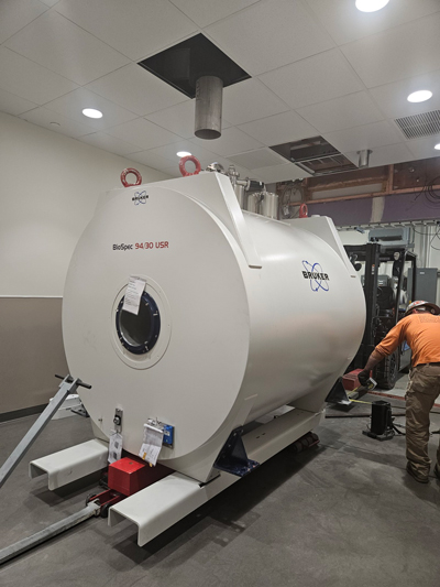
Read More: Researchers find neurons work as a team to process social interactionsThe 9.4T Bruker animal MRI scanner with PET insert is here
Saturday, October 28, 2023

2023 GEPA Award Recipients
Thursday, October 19, 2023
Several of the NGP students were recently selected for a number of the annual GEPA awards. Historically, our students have done quite well and this year is no exception. Four of our students were in the mix again laying claim to four of the awards with monetary prizes ranging from $500 - $1000. If you run into Aiesha, Amelia, Nicole, or Linh, please congratulate them, or better yet send them an email on a job well done and for keeping NGP in the spotlight. Here are a list of the awards:
| Student Award Recipients |
| Aiesha Anchan |
Graduate Alumni Fellowship Award |
| Amelia Hines |
Merritt and Marjorie Cleveland Fellowship |
| Nicole Popp |
Irving L. Spar Fellowship Award |
| Linh Le |
Outstanding Student Mentor Award |
4 NGP Faculty are also receiving Faculty and Staff Teaching Mentoring Awards as part of the GEPA awards this year. When you see any of these outstanding faculty members, make sure to also congratulate them on these recognitions. Without them, our NGP students would not be as exveptional for all these years.
| Faculty and Staff Teaching & Mentoring Award Recipients |
| Ania Majewska |
Graduate Student Society Advocacy Award |
| Nathan Smith |
Graduate Student Society Advocacy Award |
| Marissa Sobolewski |
Outstanding Graduate Student Teacher Award |
| Pat White |
Outstanding T32 Program Director Award |
These awards will be presented at the Graduate Education and Postdoctoral Affairs Awards and Philosophy Meeting on Monday, October 30, 2023 at 3:00 pm in the Flaum Atrium. We hope you make an effort to attend to support our students as well as the others receiving awards.
URMC Selected for NIH Initiative to Map Connections in the Brain
Tuesday, September 26, 2023
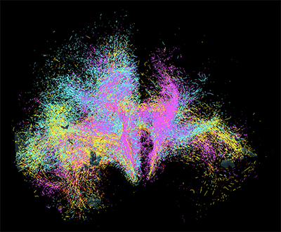 Goal is to Develop Cutting-Edge Tools to Image Neural Networks in Unprecedented Detail
Scientists at the University of Rochester Medical Center are joining research teams across the globe to develop next-generation tools for visualizing connections in the human brain. Imaging and understanding the brain’s intricate circuitry at the cellular and microscopic level will advance new approaches to treat brain disorders like Tourette’s syndrome, Parkinson’s disease, and obsessive-compulsive disorder.
The research is supported by the National Institutes of Health’s Brain Research Through Advancing Innovative Neurotechnologies® Initiative, or The BRAIN Initiative®. Principal investigator Suzanne Haber, PhD, professor of Pharmacology and Physiology at URMC, will lead a team of scientists with co-principal investigators Anastasia Yendiki of Massachusetts General Hospital and Elizabeth Hillman of Columbia University.
Haber’s lab will launch the team’s efforts by providing the “ground truth”—animal tracing data that is considered the gold standard for understanding the complex connectivity of the human brain.
“We can’t develop tools without a guide to start with, just like you can’t navigate the ocean without a map,” noted Haber, who is also a professor of Neuroscience, Brain and Cognitive Science, and Psychiatry. “Once we identify how axons traverse the brain in animal models, we use that information to develop tools to accurately track connections in the human brain. These novel technologies for imaging axonal projections will increase our understanding of the circuitry and cellular mechanisms that underlie brain abnormalities.”
Today, diffusion MRI (dMRI) is used to examine the connectivity and functional networks in the human brain. However, this is an indirect method with inherent problems, particularly in specific situations that make it difficult to accurately follow the axons. This leads to many false positives and false negatives.
 Haber says the time is right for this initiative. “We’ve seen so many exciting developments in optics and engineering over the past few years…the field has really exploded,” she notes. “We’re excited to be part of the team that’s pushing the boundaries to better understand the circuitry in humans. Ultimately, we hope this work sets the stage for the creation of more accurate diagnostics and therapeutics for brain diseases.”
Haber’s work, which will be funded by $1.5 million from NIH over five years, is part of the center for Large-scale Imaging of Neural Circuits (LINC). In addition to the University of Rochester, Massachusetts General Hospital and Columbia University, the LINC project includes collaborators from Brigham and Women’s Hospital, Harvard University, Massachusetts Institute of Technology, University College London, and Weill Cornell Medicine.
The BRAIN Initiative is a partnership between federal and non-federal partners, with the NIH taking the leading role in achieving the goal of revolutionizing our understanding of the human brain by accelerating the development of innovative neurotechnologies. The program is made up and managed by 10 NIH institutes and centers whose missions and current research portfolios complement the goals of the BRAIN Initiative. The initial round of awards is projected to total $150 million over 5 years, with a large portion of the funds dedicated to tool development.
Biomedical engineering student explores how brain processes speech syntax
Wednesday, August 16, 2023
A biomedical engineering student explores how the brain processes speech syntax—and discovers the benefits of conducting research as an undergraduate.
In a laboratory at the University of Rochester’s Center for Advanced Brain Imaging & Neurophysiology, Sophea Urbi Biswas ’24 pores over brain wave signals recorded from a person listening to an audiobook next door. Biswas, a senior biomedical engineering student from Bangladesh, is attempting to see if the syntactic features of the words and phrases the participant listens to are reflected in the waves picked up by the electroencephalography (EEG) cap they wear.
Finding such correlations is no small feat considering every blink, yawn, and head movement by the participant—not to mention all the electronic devices in the room—add noise to the experiment, making Biswas’s job harder. But she seems to relish the opportunity to apply her coding skills in a real-life setting.
“We take all these classes where they teach you the fundamentals of coding and how to use coding languages. But until now, I haven’t gotten to apply it to real data taken from real people,” says Biswas. “It’s exciting pursuing questions that have never been asked before and getting results that have never been found before. This project shows how powerful coding can be.”
Biswas earned a Schwartz Discover Grant that allowed her to spend the summer in an immersive, full-time research experience to enhance her competitiveness for future fellowships and other advanced research opportunities. After taking a class on biosystems and circuits taught by Edmund Lalor, an associate professor in the Department of Biomedical Engineering and Department of Neuroscience, they had a conversation about the research he conducts and the projects his lab was planning, and he invited her to join his research team.
Read More: Biomedical engineering student explores how brain processes speech syntaxDebra Cory-Slechta Explains How Wildfire Smoke and Air Pollution can Harm the Brain
Thursday, August 3, 2023
In an interview with Rick Woychik, director of the National Institute of Environmental Health Sciences, the professor of Environmental Medicine shares how research into how fine air particles impact the brain is transforming our understanding of neurodegenerative and neurodevelopmental disorders. Woychik interviewed Cory-Slechta after seeing her presentation at the first Human Health and the Environment Research Symposium at the Medical Center in June, where he was a guest speaker.
Read More: Debra Cory-Slechta Explains How Wildfire Smoke and Air Pollution can Harm the BrainURMC researcher receives $8.3 million to study chronic pain and the brain
Wednesday, July 19, 2023
Understanding the role of chronic pain in the brain could transform treatment and care for a condition that inflicts more than 20 percent of US adults. Paul Geha, MD, associate professor of Psychiatry has been studying the correlation between brain structure and chronic pain and was recently awarded $8.3 million from the National Institute of Neurological Disorders and Stroke (NINDS) and the National Institute of Arthritis and Musculoskeletal and Skin Diseases (NIAMS) to continue this work in understanding the link between pain and the brain.
His previous research found a possible explanation to why people with chronic pain often struggle with their weight. The research published in PLOS ONE suggests that circuitry in the brain responsible for motivation and pleasure is impacted when someone experiences pain, changing their brain’s response to fat and brain scans showed disrupted satiety signals.
“Our ongoing research focuses on unraveling the mechanisms underlying pain perception in the brain, as well as utilizing brain circuitries to gain insights into the pathophysiology of chronic pain. Additionally, we aim to develop quantitative biomarkers that can be derived from natural language processing of patients' pain narratives and multimodal brain imaging,” said Geha. “These biomarkers hold the potential to predict clinical outcomes in patients, including response to analgesics or the long-term persistence or remission of pain.”
Read More: URMC researcher receives $8.3 million to study chronic pain and the brainImages capture unseen details of the synapse
Wednesday, June 14, 2023
Scientists have created one of the most detailed 3D images of the synapse, the important juncture where neurons communicate with each other through an exchange of chemical signals. These nanometer scale models will help scientists better understand and study neurodegenerative diseases such as Huntington’s disease and schizophrenia.
The new study appears in the journal PNAS and was authored by a team led by Steve Goldman, MD, PhD, co-director of the Center for Translational Neuromedicine at the University of Rochester and the University of Copenhagen. The findings represent a significant technical achievement that allows researchers to study the different cells that converge at individual synapses at a level of detail not previously achievable.
“It is one thing to understand the structure of the synapse from the literature, but it is another to see the precise geometry of interactions between individual cells with your own eyes,” said Abdellatif Benraiss, PhD, a research associate professor in the Center for Translational Neuromedicine and co-author of the study. “The ability to measure these extremely small environments is a young field, and holds the potential to advance our understanding of a number of neurodegenerative and neuropsychiatric diseases in which synaptic function is disturbed.”
Read More: Images capture unseen details of the synapseA chance observation finds potential hearing biomarker for Alzheimer’s disease
Wednesday, June 7, 2023
Science lends itself to questions, changing hypotheses, and chance findings. Recently, in the White Lab at the Del Monte Institute for Neuroscience at the University of Rochester, Neuroscience graduate student Daxiang Na was reviewing data for one project but instead uncovered something unexpected. He discovered that where plaques associated with Alzheimer’s disease are found in the brain may contribute to hearing loss.
Na was conducting hearing tests on mice with amyloid beta, the main component of protein plaques and tangles found in Alzheimer’s. While looking at two different transgenic mouse models of the disease, he found for one model, called 5xFAD, the older mice had hearing changes similar to what is found in people with Alzheimer’s disease. The other model did not demonstrate these hearing changes, nor did younger mice in the 5xFAD group.
“It was a chance observation,” said Na, who is first author of a paper with these findings in Frontiers in Neuroscience. “Both mouse models had amyloid beta protein, but where we found the plaque varied, and that may be why hearing loss varied across the groups.”
Researchers found that the brains of older mice from both models had plaques in the hippocampus and auditory cortex. But the brain of mice with hearing changes also had a small amount of plaque on the auditory brainstem, suggesting this area may be sensitive to disruption from plaque found in Alzheimer’s. Researchers discovered that the plaque reduced the brainstem’s ability to coordinate responses to sound.
“This may explain why Alzheimer’s patients have auditory symptoms,” said Patricia White, PhD, professor of Neuroscience and senior author of the study. “We think the location of plaques may be more important to hearing decline. It could be a potential biomarker to track disease progression because it could be assessed with amyloid PET imaging. Our data also suggest that regular auditory Brainstem Response assessments could help with diagnosis.”
Read More: A chance observation finds potential hearing biomarker for Alzheimer’s diseaseResearch finds prediction may be key to eye-and-hand coordination
Monday, June 5, 2023
Have you ever made a great catch—like saving a phone from dropping into a toilet or catching an indoor cat from running outside? Those skills—the ability to grab a moving object—takes precise interactions within and between our visual and motor systems. Researchers at the Del Monte Institute for Neuroscience at the University of Rochester have found that the ability to visually predict movement may be an important part of the ability to make a great catch—or grab a moving object.
“We were able to develop a method that allowed us to analyze behaviors in a natural environment with high precision, which is important because, as we showed, behavioral patterns differ in a controlled setting,” said Kuan Hong Wang, PhD, a Dean’s Professor of Neuroscience at the University of Rochester Medical Center. Wang co-led the study out today in Current Biology with Jude Mitchell, PhD, assistant professor of Brain and Cognitive Sciences at the University of Rochester. “Understanding how natural behaviors work will give us better insight into what is going awry in an array of neurological disorders.”
Read More: Research finds prediction may be key to eye-and-hand coordinationResearchers find possible target for treating neuropsychiatric disorders in teens
Thursday, June 1, 2023
The brain continuously changes during childhood and throughout adolescence. The onset of neuropsychiatric disorders like schizophrenia often begins during young adulthood. Dysfunction of the dopamine system—necessary for cognitive processing and decision-making—begins during this point in development. Researchers at the Del Monte Institute for Neuroscience at the University of Rochester are coming closer to finding a possible target for treating neuropsychiatric disorders like schizophrenia and autism during this time of development that could affect the brain circuitry into adulthood.
“Brain development is a lengthy process, and many neuronal systems have critical windows—key times when brain areas are malleable and undergoing final maturation steps,” said Rianne Stowell, PhD, a postdoctoral fellow in the Wang Lab at the University of Rochester Medical Center and co-first author on research out today in the journal eLife. “By identifying these windows, we can target interventions to these time periods and possibly change the course of a disease by rescuing the structural and behavioral deficits caused by these disorders.”
Read More: Researchers find possible target for treating neuropsychiatric disorders in teensResearchers: Early alcohol exposure does not change connection between brain’s immune system and neurons that send information related to functions like balance and memory (UPDATE)
Monday, May 8, 2023
Research out of the Majewska Lab at the Del Monte Institute for Neuroscience at the University of Rochester continues to show the brain’s immune system does not play a significant role in the neurological damage that occurs in fetal alcohol spectrum disorders (FASD).
A new study out in Frontiers in Neuroscience investigated the interaction between microglia and Purkinje neurons—the neurons responsible for sending information from the cerebellum. Researchers found mice exposed to ethanol during development had no differences in microglia movement or structure and only subtle changes in the interaction between microglia and Purkinje neurons. “It appears that developmental ethanol exposure has little effect on microglia later in life,” said MaKenna Cealie, a Neuroscience graduate student in the lab and first author of the paper. “We believe examining other cell types and their interactions may be an important direction for future FASD studies to take.”
Other authors include the senior author Ania Majewska, PhD, Linh Le, Erik Vonkaenel, and Matthew McCall, PhD, of the University of Rochester Medical Center, and James Douglas and Paul Drew, PhD, of the University of Arkansas Medical Center.
The research was supported by the National Institutes of Health (NIH), the National Institute on Alcohol Abuse and Alcoholism (NIAAA), and the University of Rochester Intellectual and Developmental Disabilities Research Center (UR-IDDRC).
Read More: Researchers: Early alcohol exposure does not change connection between brain’s immune system and neurons that send information related to functions like balance and memory (UPDATE)Halting the Rise of Parkinson’s
Monday, April 24, 2023
Quality of life, health, and longevity are being increasingly tied to someone’s zip code rather than their genetic code. Cancer, heart disease, neurodegenerative disorders, and even our ability to fight infection are linked to the myriad of chemicals we are exposed to, often unwittingly, over the course of our lives. The University of Rochester’s leadership in the field of environmental medicine stretches back to toxicology research programs developed at the University under the Manhattan Project. These programs also served as the basis for the formation of a NIEHS Center of Excellence in environmental toxicology and health that is one of the oldest in the country celebrating 50 years of sustained funding. This foundation and the decades of work that followed—coupled with the recognition that the public health threat requires a collaborative commitment to research, education, and community engagement—led to the creation of the new Institute for Human Health and the Environment.
Paige Lawrence, PhD, the Wright Family Research Professor and chair of the Department of Environmental Medicine, is the founding director of the new Institute. “Genetics only explaining 10 to 15 percent of human health, which leaves the rest to the environment,” said Lawrence. “If we really want to have an impact on health, environmental influences need to be front and center.”
The new Institute will help power a team of neurologists, neuroscientists, toxicologists, epidemiologists, and researchers at the University of Rochester who are examining the impact of environmental chemical exposure on the brain. One disease in particular stands out. Parkinson’s is the fastest growing neurodegenerative disease in the world, outpacing even Alzheimer’s, and a growing number of scientists are linking the disease’s rise to air pollution, pesticides, and a ubiquitous chemical pollutant.
Up the nose it goes
Air pollution is associated with many health problems, including asthma, heart disease, stroke, low birth weight, and inflammation. While epidemiological studies have hinted at the link between air pollution and neurological disorders like Parkinson’s and Alzheimer’s, the route these chemicals use to make their way into the brain, and the damage caused once there, was until recently poorly understood.
“We’ve known that air pollution has effects on the heart and the lung for a very long time, but it's really only been in about the past ten years that attention has been directed to its effects on the brain,” said Debbie Cory-Slechta, PhD, a professor of Environmental Medicine, Neuroscience, and Public Health Sciences. Cory-Slechta’s colleagues at the University of Rochester, Guenter Oberdoerster, PhD, and Alison Elder, PhD, were among the first to show that ultra-fine air pollution particles, called PM0.1, are able to hitch a ride directly into the brain via the nasal passage and olfactory nerves, bypassing the brain’s normal defenses.
Read More: Halting the Rise of Parkinson’sIan Fiebelkorn finds rhythmic brain activity helps to maintain temporary memories
Monday, April 24, 2023
New research shows that rhythmic brain activity is key to temporarily maintaining important information in memory. Researchers at the Del Monte Institute for Neuroscience at the University of Rochester published these findings today in Current Biology that found brain rhythms—or patterns of neuronal activity—organize the bursts of activity in the brain that maintain short-term connections.
“The thought has been that the temporary storage of important information is linked to neurons in the brain that just fire away, retaining that information until it is no longer needed. Recent research has shown that it might not be such persistent brain activity that matters most for the temporary storage of information, but rather a short-term strengthening of the connections between neurons that are representing the information. Our research shows that brain rhythms are organizing these transient bursts over time,” said Ian Fiebelkorn, PhD, assistant professor of Neuroscience and senior author of the study. “The rhythmic coordination of brain activity over time is important because it allows overlapping populations of neurons to store different pieces of information at the same time.”
Fiebelkorn’s previous research around how the brain processes external information—like when navigating Times Square in New York City—made a similar discovery. He and fellow researchers found that brain rhythms help to coordinate different functions associated with either sampling presently important information or shifting to another source of information. In this context, brain rhythms help to balance focus on the task at hand with being prepared for the unexpected.
Read More: Ian Fiebelkorn finds rhythmic brain activity helps to maintain temporary memoriesPossible ‘steps’ to revealing super-agers
Thursday, April 13, 2023
On the quest for the proverbial fountain of youth, scientists have long looked for evidence of super-agers—people whose brain ages slower than their body. Researchers at the Del Monte Institute for Neuroscience at the University of Rochester have found older adults whose brain performance improves when they combine a cognitive task with walking.
“Identifying super-agers will leverage what we understand about the brain and aging,” said Eleni Patelaki, a Biomedical Engineering PhD student at the University of Rochester Medical Center and first author of the paper out today in NeuroImage. “But this is difficult to do because, in this case, there was no external evidence of this ability, and people are unaware that their brain is working differently.
Walking and doing exposes brain flexibility
Researchers had the participants complete the same cognitive task while sitting and while walking. The 37 men and women, ages 62 to 79, scored similarly while sitting. When the same group repeated the test while walking, researchers found some individuals improved their cognitive performance. Researchers used Mobile Brain/Body Imaging (MoBI) to observe these changes and measure how the brain responded to the dual task. “We think this brain activity might constitute signatures of ‘super-aging,” said Patelaki. “We were able to find seven people, and now that we know where and how to look in the brain to find these super-agers, we can find more.”
The participants whose cognition improved while walking showed that their brain was able to adapt to and improve at the task—it had flexible usage of certain frontal resources. But those same people lost their flexibility in using the rest of their neural resources, similar to their peers who did not improve at the task while walking. This suggests that the brain’s ability to adapt or its flexibility in reallocating neural resources while walking might be an important factor in protecting cognition as we age.
Read More: Possible ‘steps’ to revealing super-agersThe stars in the brain may be information regulators
Friday, March 31, 2023
URMC researcher, Nathan A. Smith,MS, PhD, explores how astrocytes may be a key player in the brain’s ability to process external and internal information simultaneously. He argues research on these cells is necessary to understand their role in the process that allows a person to have an appropriate behavioral response and also the ability to create a relevant memory to guide future behavior.
Read More: The stars in the brain may be information regulatorsIron & the brain: Where and when neurodevelopmental disabilities may begin during pregnancy
Monday, March 6, 2023
Study finds possible cellular origin for impairments associated with gestational iron deficiency
The cells that make up the human brain begin developing long before the physical shape of the brain has formed. This early organizing of a network of cells plays a major role in brain health throughout the course of a lifetime. Numerous studies have found that mothers with low iron levels during pregnancy have a higher risk of giving birth to a child that develops cognitive impairments like autism, attention deficit syndrome, and learning disabilities. However, iron deficiency is still prevalent in pregnant mothers and young children.
The mechanisms by which gestational iron deficiency (GID) contributes to cognitive impairment are not fully understood. The laboratory of Margot Mayer- Proschel, PhD, a professor of Biomedical Genetics and Neuroscience at the University of Rochester Medical Center, was the first to demonstrated that the brains of animals born to iron-deficient mice react abnormally to excitatory brain stimuli, and that iron supplements giving at birth does not restore functional impairment that appears later in life. Most recently, her lab has made a significant progress in the quest to find the cellular origin of the impairment and have identified a new embryonic neuronal progenitor cell target for GID. This study was recently published in the journal Development.
“We are very excited by this finding,” Mayer-Proschel said, who was awarded a $2 million grant from the National Institute of Child Health & Human Development in 2018 to do this work. “This could connect gestational iron deficiency to these very complex disorders. Understanding that connection could lead to changes to healthcare recommendations and potential targets for future therapies.”
Read More: Iron & the brain: Where and when neurodevelopmental disabilities may begin during pregnancyThrough the eye of the beholder: People with autism may process illusory shapes differently
Tuesday, February 28, 2023
Researchers are finding the process in our brain that allows us to see these visual distinctions may not be happening the same way in the brains of children with autism spectrum disorder. They may be seeing these illusions differently.
 There is this picture – you may have seen it. It is black and white and has two silhouettes facing one another. Or maybe you see the black vase with a white background. But now, you likely see both.
It is an example of a visual illusion that reminds us to consider what we did not see at first glance, what we may not be able to see, or what our experience has taught us to know – there is always more to the picture or maybe even a different image to consider altogether. Researchers are finding the process in our brain that allows us to see these visual distinctions may not be happening the same way in the brains of children with autism spectrum disorder. They may be seeing these illusions differently.
 “How our brain puts together pieces of an object or visual scene is important in helping us interact with our environments,” said Emily Knight, MD, PhD, assistant professor of Neuroscience and Pediatrics at the University of Rochester Medical Center, and first author on a study out today in the Journal of Neuroscience. “When we view an object or picture, our brains use processes that consider our experience and contextual information to help anticipate sensory inputs, address ambiguity, and fill in the missing information.”
Small, involuntary eye movements help us see a stable world
Thursday, February 23, 2023
Our eyes are never at rest. Instead, they remain in motion, even between our voluntary gaze shifts, through fixational eye movements—small, continuous movements of the eye that we are not aware of making.
Scientists have long sought to understand how we humans can perceive the world as stable as our eyes are constantly moving. Past research has suggested that, in the intervals between voluntary gaze shifts, the human visual system builds a picture of a stable world by relying solely on sensory inputs from fixational eye movements. According to new research by a team at Rochester, however, there may be another contributing factor.
In a paper published in Nature Communications, the researchers—including Michele Rucci, a professor of brain and cognitive sciences and, and first author Zhetuo Zhao, a PhD student in Rucci’s lab—report that the visual system not only receives sensory inputs from fixational eye movements but also possesses knowledge of the motor behavior involved in those movements.
Read More: Small, involuntary eye movements help us see a stable worldCan hearing loss be reversed? Research in Patricia White's lab reveals clues that could regrow the cells that help us hear
Monday, February 13, 2023
Taking a bite of an apple is considered a healthy choice. But have you ever thought about putting in earplugs before your favorite band takes the stage?
Just like your future body will thank you for the apple, your future ears (specifically your cochlear hair cells) will thank you for protecting them. The most common cause of hearing loss is progressive because these hair cells—the primary cells to detect sound waves—cannot regenerate if damaged or lost. People who have repeated exposure to loud noises, like military personnel, construction workers, and musicians, are most at risk for this type of hearing loss. But, it can happen to anyone over time (even concert goers).
On the other hand, birds and fish can regenerate these hair cells, and now researchers at the Del Monte Institute for Neuroscience are getting closer to identifying the mechanisms that may promote this type of regeneration in mammals, as explained in research recently published in Frontiers in Cellular Neuroscience.
 “We know from our previous work that expression of an active growth gene, called ERBB2, was able to activate the growth of new hair cells (in mammals), but we didn’t fully understand why,” said Patricia White, PhD, professor of Neuroscience and Otolaryngology at the University of Rochester Medical Center. The 2018 study led by Jingyuan Zhang, PhD, a postdoctoral fellow in the White lab at the time, found that activating the growth gene ERBB2 pathway triggered a cascading series of cellular events by which cochlear support cells began to multiply and activate other neighboring stem cells to become new sensory hair cells.
Read More: Can hearing loss be reversed? Research in Patricia White's lab reveals clues that could regrow the cells that help us hearResearchers identify neurons that "learn" to smell a threat
Tuesday, January 24, 2023
Whether conscious of it or not, when entering a new space, we use our sense of smell to assess whether it is safe or a threat. In fact, for much of the animal kingdom, this ability is necessary for survival and reproduction. Researchers at the Del Monte Institute for Neuroscience at the University of Rochester are finding new clues to how the olfactory sensory system aids in threat assessment and have found neurons that “learn” if a smell is a threat.

Julian Meeks, PhD
“We are trying to understand how animals interact with smell and how that influences their behavior in threatening social and non-social contexts,” said Julian Meeks, PhD, principal investigator of the Chemosensation and Social Learning Laboratory. “Our recent research gives us valuable tools to use in our future work and connects specific sets of neurons in our olfactory system to the memory of threatening smells.”
Read More: Researchers identify neurons that "learn" to smell a threatKerry O'Banion speaks with Medical News Today
Wednesday, January 18, 2023
Gut-brain connection: 3 fatty acids may be linked to tau-mediated damage
As the prevalence of Alzheimer’s disease (AD) continues to increase, the search for ways to treat and prevent it is ever more pressing. Newly licensed treatments, such as aducanumab and lecanemab, that clear beta-amyloidTrusted Source from the brain are a positive development, but they are expensive and controversial.
Many researchers are now focusing on other areas, one of which is the effect of the microbiomeTrusted Source — microbes, particularly bacteria, that inhabit the gut — on neurodegenerative disorders.
. "There is growing recognition of a gut-brain axis and evidence that the microbiome of individuals varies with disease status," said O'Banion. "The biggest issue is understanding whether gut changes are due to disease or contribute to disease (or both)."
Read More: Kerry O'Banion speaks with Medical News Today