Anatomy of the Uterus
The uterus is an organ in the lower belly (abdomen) or pelvis. It is part of the female
reproductive system. It's where a baby grows. It's sometimes called the womb.
The uterus is hollow and pear-shaped. It is about the size of a fist. It's in your
lower belly (pelvic area). Your uterus is connected to the fallopian tubes. These
tubes help carry eggs from the ovaries into the uterus. The lower part of the uterus
connects to the vagina and is called the cervix. The wider, upper part of the uterus
is called the corpus or fundus.
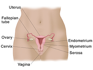
The uterus has 3 layers:
-
Endometrium. This is the inner lining. It's shed during a menstrual period.
-
Myometrium. This is the thick middle muscle layer of the corpus or fundus. It expands during pregnancy
to hold the growing baby. It contracts during labor to push the baby out.
-
Serosa. This is the smooth outer layer. It covers the uterus and makes it easy for the uterus
to slide and move within the pelvis as needed.
In people who still have their periods, 1 ovary releases an egg into a fallopian tube
each month. During this time, the endometrium becomes thicker to prepare for a fertilized
egg. The egg enters the uterus. If it isn’t fertilized, it leaves the uterus through
the vagina, and the endometrial lining is shed during a menstrual period. If the egg
joins with a sperm cell (from a male), this fertilized egg attaches to the endometrium.
The thick wall of the uterus protects the growing baby during pregnancy. During labor,
the cervix opens (dilates). The muscles of the myometrium help push the baby out through
the vagina.
The balance of the female hormones estrogen and progesterone control this process.
Most of these hormones are made by the ovaries.