Endoscopic Exam for Cancer
How is cancer diagnosed?
There isn't 1 test that can accurately diagnose cancer. A full medical history and
physical exam along with diagnostic testing is often needed.
Many tests are used to find out if a person has cancer, or if another condition (such
as an infection) is causing symptoms that are like those caused by cancer. Diagnostic
testing is used to confirm or rule out cancer, watch cancer growth, plan for treatment,
and see if treatment is working. In some cases, repeat testing is needed when your
condition has changed. Repeat testing is also needed if a sample collected was not
of good quality, or an abnormal test result needs to be confirmed. Diagnostic procedures
for cancer include imaging, lab tests (including blood tests for tumor markers), tumor
biopsy, endoscopic exam, surgery, and genetic testing.
Endoscopic exams are used to look inside the body using a thin, tube-like device called
an endoscope. These exams are covered here.
What are some of the different types of endoscopic exams?
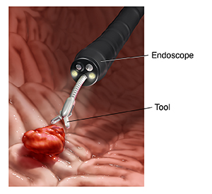
An endoscope is a small, flexible tube with a light and a lens or tiny video camera
on the end. It can be used to look into the esophagus, stomach, duodenum, colon, rectum,
or other organs. It can also be used to take tissue from the body for testing (called
a biopsy). Or it can be used to take color photos of the inside of the body. Different
types of endoscopes that are often used to diagnose cancer include:
-
Colonoscopy. This procedure allows the healthcare provider to see the entire length of the large
intestine (colon). It can often help find abnormal growths, inflamed tissue, ulcers,
and bleeding. A long, flexible, lighted tube (colonoscope) is put in through the rectum
up into the colon. This scope lets the provider see the lining of the colon, remove
tissue for further exam, and possibly treat some problems that are found.
-
ERCP (endoscopic retrograde cholangiopancreatography). An ERCP allows the healthcare provider to diagnose and treat problems in the liver,
gallbladder, bile ducts, and pancreas. The procedure combines X-ray and the use of
an endoscope. The scope is guided through the person's mouth and throat, then through
the esophagus, stomach, and the first part of the small intestine (the duodenum).
The provider can check the inside of these organs and find any abnormalities. A tube
is then passed through the scope and a dye is injected into the pancreatic duct. This lets
the internal organs be evaluated using X-rays. This will allow the internal organs
to be seen more clearly on an X-ray.
-
EGD or upper endoscopy (esophagogastroduodenoscopy). This procedure allows the healthcare provider to examine the inside of the esophagus,
stomach, and duodenum. The endoscope is guided into the mouth and throat. It then
goes into the esophagus, stomach, and duodenum. The endoscope lets the provider see the
inside of this part of the body. The provider can also insert tiny tools through the
scope to remove a tissue sample for biopsy, if needed.
-
Sigmoidoscopy. This diagnostic procedure lets the healthcare provider examine the inside of a part of
the large intestine. It can be helpful in identifying the causes of diarrhea, stomach
pain, constipation, abnormal growths, and bleeding. This procedure uses a short, flexible,
lighted tube called a sigmoidoscope. It is put into the intestine through the rectum.
The scope blows air into the intestine to inflate it and make viewing the inside easier.
-
Bronchoscopy. This diagnostic procedure allows the provider to examine the inside of the windpipe
(trachea) and large airways leading into the lungs (bronchi). A short, flexible, lighted
tube (bronchoscope) is inserted through the mouth or nose. Tissue samples may be removed
through the bronchoscope for exam under a microscope in the lab.
-
Cystoscopy. For this exam, a flexible tube with a viewing device (cystoscope) is inserted through
the urethra. It is used to check the bladder and urinary tract for structural abnormalities
or blockages, such as tumors or stones. Samples of the bladder tissue may be removed
through the cystoscope for exam under a microscope in the lab.
-
Laryngoscopy. For this exam, the healthcare provider uses a thin, flexible tube with a light and
magnifier (laryngoscope) to see the back of your throat, voice box, and vocal cords.
Your healthcare provider can tell you more about a specific test. This includes where
it will be done, how to get ready, and what recovery will look like.