External and Internal Heart Rate Monitoring of the Fetus*
(Fetal Monitoring, External and Internal)
Procedure Overview
What is external and internal fetal heart rate monitoring?
Fetal heart rate monitoring is a procedure used to evaluate the well-being of the
fetus by assessing the rate and rhythm of the fetal heartbeat.
During late pregnancy and labor, your physician may recommend monitoring the fetal
heart rate and other functions. The average fetal heart rate is between 110 and 160
beats per minute, and can vary five to 25 beats per minute. The fetal heart rate may
change as the fetus responds to conditions in the uterus. An abnormal fetal heart
rate or pattern may indicate that the fetus is not getting enough oxygen or that there
are other problems.
There are two methods for fetal heart rate monitoring, external and internal:
-
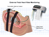
External fetal heart rate monitoring uses a device to listen to or record the fetal
heartbeat through the mother's abdomen. A fetoscope (a type of stethoscope) is the
most basic type of external monitor. Another type of monitor is a hand-held electronic
Doppler ultrasound device. These methods are often used during prenatal visits to
count the fetal heart rate. A fetoscope or Doppler device may also be used to check
the fetal heart rate at regular intervals during labor.Continuous electronic fetal
heart monitoring may be used during labor and birth. An ultrasound transducer placed
on the mother's abdomen conducts the sounds of the fetal heart to a computer. The
rate and pattern of the fetal heart are displayed on the computer screen and printed
onto special graph paper.
-
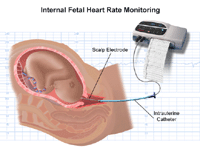
Internal fetal heart rate monitoring uses an electronic transducer connected directly
to the fetal skin. A wire electrode is attached to the fetal scalp or other body part
through the cervical opening and is connected to the monitor. This type of electrode
is sometimes called a spiral or scalp electrode. Internal monitoring provides a more
accurate and consistent transmission of the fetal heart rate than external monitoring
because factors such as movement do not affect it. Internal monitoring may be used
when external monitoring of the fetal heart rate is inadequate, or closer surveillance
is needed.
During labor, uterine contractions are usually monitored along with the fetal heart
rate. A pressure-sensitive device called a tocodynamometer is placed on the mother's
abdomen over the area of strongest contractions to measure the length, frequency,
and strength of uterine contractions. Because the fetal heart rate and uterine contractions
are recorded at the same time, these results can be examined together and compared.
Internal uterine pressure monitoring is sometimes used along with internal fetal heart
rate monitoring. A fluid-filled catheter is placed through the cervical opening into
the uterus beside the fetus and transmits uterine pressure readings to the monitor.
Other procedures that may be used to monitor the well-being of the fetus include amniocentesis
and chorionic villus sampling. Please see these procedures for additional information.
Anatomy of the fetus:

-
amniotic sac - a thin-walled sac that surrounds the fetus during pregnancy. The sac is filled
with amniotic fluid (liquid made by the fetus) and the amnion (the membrane that covers
the fetal side of the placenta), which protects the fetus from injury and helps to
regulate the temperature of the fetus.
-
anus - the opening at the end of the anal canal
-
cervix - the lower part of the uterus that projects into the vagina. Made up of mostly fibrous
tissue and muscle, the cervix is circular in shape.
-
fetus - an unborn baby from the eighth week after fertilization until birth
-
placenta - an organ, shaped like a flat cake, that only grows during pregnancy and provides
a metabolic interchange between the fetus and mother. (The fetus takes in oxygen,
food, and other substances and eliminates carbon dioxide and other wastes.)
-
umbilical cord - a rope-like cord connecting the fetus to the placenta. The umbilical cord contains
two arteries and a vein, which carry oxygen and nutrients to the fetus and waste products
away from the fetus.
-
uterine wall - the wall of the uterus
-
uterus (Also called the womb.) - the uterus is a hollow, pear-shaped organ located in a woman's lower abdomen, between
the bladder and the rectum, that sheds its lining each month during menstruation and
in which a fertilized egg (ovum) becomes implanted and the fetus develops
-
vagina - the part of the female genitals, behind the bladder and in front of the rectum,
that forms a canal extending from the uterus to the vulva
Reasons for the Procedure
Fetal heart rate monitoring is used in nearly every pregnancy to assess fetal well-being
and identify any changes that might be associated with problems during pregnancy or
labor. Fetal heart rate monitoring is especially helpful for high-risk pregnancy conditions
such as diabetes, high blood pressure, and problems with fetal growth.
Situations during pregnancy in which fetal heart rate monitoring may be used include,
but are not limited to, assessment of fetal heart rate during prenatal physician visits
and monitoring the effect of preterm labor medications on the fetus.
Fetal heart rate monitoring may be used as a component of other procedures, including,
but not limited to, the following:
-
nonstress test (a procedure that measures the fetal heart rate in response to fetal
movements)
-
a contraction stress test (a procedure in which the fetal heart rate is observed with
uterine contractions which have been stimulated with medication or other methods)
-
a biophysical profile, or BPP (a test that combines a nonstress test with ultrasound)
Situations during labor which may affect the fetal heart rate and for which fetal
heart rate monitoring may be used include, but are not limited to, the following:
-
uterine contractions
-
pain medications and/or anesthetic agents given to the mother during labor
-
procedures performed during labor
-
pushing during the second stage of labor
There may be other reasons for your physician to recommend fetal heart rate monitoring.
Risks of the Procedure
There is no radiation used and generally no discomfort from the application of the
transducer to the abdominal skin.
The elastic belts that hold the ultrasound and pressure transducers in place around
your abdomen may be slightly uncomfortable. These can be readjusted to help you feel
more comfortable.
You must lie still during some types of fetal heart rate monitoring. You may be required
to stay in bed during labor.
With internal monitoring, you may experience some slight discomfort during the insertion
of the electrode.
Risks of internal monitoring include, but are not limited to, infection and bruising
of the fetal scalp or other body part.
Internal fetal heart rate monitoring is contraindicated in women with active herpes
lesions on the cervix or vagina because of the risk of transferring the infection
to the fetus.
There may be other risks depending upon your specific medical condition. Be sure to
discuss any concerns with your physician prior to the procedure.
Certain factors or conditions may interfere with the results of the procedure. These
include, but are not limited to, the following:
-
obesity of the mother
-
position of the fetus or mother
-
too much amniotic fluid (polyhydramnios)
-
cervix not adequately dilated or amniotic sac not broken (for internal monitoring)
Before the Procedure
-
Your physician will explain the procedure to you and offer you the opportunity to
ask any questions that you might have about the procedure.
-
You may be asked to sign a consent form that gives your permission to do the procedure.
Read the form carefully and ask questions if something is not clear. The consent form
for fetal heart rate monitoring may be included as part of the general consent for
your labor and birth.
-
If fetal heart rate monitoring is performed in conjunction with another monitoring
procedure, such as a nonstress test or biophysical profile, you may be asked to eat
a meal before the procedure. This can help increase fetal activity. You may or may
not be able to eat or drink with fetal heart rate monitoring during labor.
-
Although the gel applied to the skin during the procedure does not stain clothing,
you may wish to wear older clothing or a hospital gown, as the gel may not be completely
removed from your skin afterwards.
-
Placement of an internal electrode requires that the amniotic membranes (the fluid-filled
sac that surrounds the fetus during pregnancy) be broken, and the cervix be dilated
(opened) several centimeters to allow contact with the fetal scalp or other body part.
-
Based upon your medical condition, your physician may request other specific preparation.
During the Procedure
Fetal heart rate monitoring may be performed in your physician's office, on an outpatient
basis, or as part of your stay in a hospital. Procedures may vary depending on your
condition and your hospital's practices.
Generally, fetal heart rate monitoring follows this process:
For external fetal heart rate monitoring:
-
Depending on the type of procedure, you may be asked to expose your abdomen, undress
from the waist down, or undress completely and put on a hospital gown.
-
You will lie on your back on an examination table.
-
A clear gel will be applied to your abdomen (the gel acts as a conductor).
-
The transducer will be pressed against the skin and moved around until the fetal heartbeat
is located. You will be able to hear the sound of the fetal heart rate with Doppler
or an electronic monitor.
-
During labor, the fetal heart rate may be monitored intermittently or continuously,
depending on your condition and the condition of your fetus.
-
For continuous electronic monitoring, the transducer will be connected to the monitor
with a cable. A wide elastic belt will be placed around your back to secure the transducer
in place.
-
The fetal heart rate will be recorded in the medical record. With continuous electronic
monitoring, the fetal heart pattern will be displayed on a computer screen and printed
onto graph paper.
-
You may or may not be allowed to get out of bed with continuous external fetal heart
rate monitoring.
-
Once the procedure has been completed, the transducer will be removed and the gel
will be wiped off.
For internal fetal heart rate monitoring:
-
You will be asked to undress completely and put on a hospital gown, and lie on a labor
bed, with your feet and legs supported as for a pelvic exam.
-
The physician or nurse will perform a vaginal examination with a gloved hand to check
cervical dilation (the amount the cervix has opened). This may be slightly uncomfortable.
-
If the amniotic sac is still intact, your physician may break open the membranes with
an instrument. You will feel warm fluid coming out of your vagina.
-
The physician will feel the part of the fetus at the cervical opening with gloved
fingers - this is usually the fetus head.
-
A long, plastic electrode guide will be inserted into your vagina. A small spiral
wire at the end of the electrode will be placed against the fetal part and gently
rotated into the fetal skin.
-
The guide will be removed and the electrode will be left in place, attached to the
fetal part.
-
The electrode wires will be connected to the monitor cable and secured with a band
around your thigh.
-
You may or may not be allowed to get out of bed with continuous internal fetal heart
rate monitoring.
-
Once the baby is born, the electrode will be removed.
After the Procedure
There is no special type of care required after external fetal heart rate monitoring.
You may resume your normal diet and activity unless your physician advises you differently.
After internal fetal heart rate monitoring, the electrode site on the newborn baby
will be examined for infection, bruising, or a laceration. The site may be cleansed
with an antiseptic.
Your physician may give you additional or alternate instructions after the procedure,
depending on your particular situation.
Online Resources
The content provided here is for informational purposes only, and was not designed
to diagnose or treat a health problem or disease, or replace the professional medical
advice you receive from your physician. Please consult your physician with any questions
or concerns you may have regarding your condition.
This page contains links to other Web sites with information about this procedure
and related health conditions. We hope you find these sites helpful, but please remember
we do not control or endorse the information presented on these Web sites, nor do
these sites endorse the information contained here.
American College of Obstetricians and Gynecologists
American Institute of Ultrasound in Medicine
National Institutes of Health (NIH)
National Library of Medicine
National Women's Health Information Center