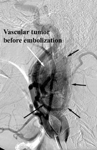
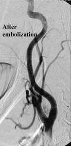
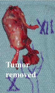
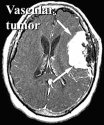
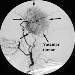
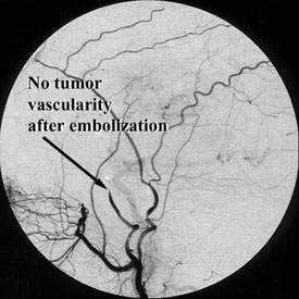
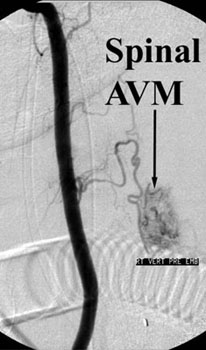
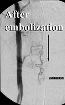
Many of the tumors that occur in the head, neck, and spine have a large blood supply. This can make surgical removal of these tumors difficult and risky. These tumors include meningiomas (tumors of the covering of the brain), paragangliomas or glomus tumors (tumors associated with nerves of the head and neck), juvenile nasopharyngeal angiofibromas (tumors of the nose that occur in young males), head and neck cancers, and tumors of the bones of the spine (vertebrae). When surgery is planned, a catheter can be placed into an artery (usually in the leg, like for an angiogram of the heart) and a tiny catheter threaded up through this to the artery or arteries supplying the tumor. Material is injected to block off the blood supply to the tumor (this is called embolization). There are many different kinds of materials available for this, depending on the type of tumor, its location, and the size of the blood vessels. This is usually performed within a few days before surgery. Sometimes, especially in the case of tumors of the vertebrae (or other bones), a needle is inserted through the skin directly into the bone containing the tumor and material is injected to block the blood supply or kill the tumor.