 A spinal angiogram is an x-ray study of the blood vessels supplying the spine. It is usually performed to look for abnormality of the vessels, so called vascular malformations. It may also be done to treat a vascular tumor or a vascular malformation.
A spinal angiogram is done in an angiography suite with an x-ray machine. You will have to have fasted for 4 to 6 hours before the procedure. There will be a doctor and nurse and a radiologic technologist in the room. The groin is anesthetized with local anesthesia and the artery in the groin is punctured and a long fine catheter is feed up into the artery. The tip of the catheter is placed in the spinal arteries. Multiple views are needed with multiple injections of each of the spinal arteries.
Spinal angiograms are often performed to evaluate for arteriovenous malformations related to the spinal cord. It can be done with MR imaging, but often conventional angiography is necessary for finer details.
Spinal angiography is a tedious process because there is one spinal artery for each rib and often you cannot tell before the study which artery is supplying the vascular malformation. During the procedure it is important to keep track of which injection represents which artery since the printed images look the same and there is no way to count on the individual images.
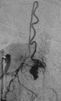
In the images there is a case where we could use MR angiography to localize which artery was feeding the arteriovenous malformation, then we would have to do a conventional spinal digital subtraction angiography.