Recognition of specific apoptotic cells by CLL antibody is critical for maintenance and evolution of CLL clone
My laboratory has been defining the molecule(s) that bind to the antibodies made by CLL cells in collaboration with Drs. Nicholas Chiorazzi, M.D. and Kanti Rai, M.D. at the Feinstein Institute for Medical Research. We have shown that antibodies from one subset of CLL patients, whose cells express an unmutated antibody characterized by a heavy (H) chain encoded by IGHV1-69, IGHD3-16, and IGHJ3 and a light (L) chain encoded by IGKV3-20, bind an intracellular protein, non-muscle myosin heavy chain IIA (myosin). We have further shown that myosin becomes exposed on the outside of the cell during programmed cell death (apoptosis), and thus becomes available to stimulate CLL cell growth. Interestingly, not all dying cells expose myosin outside of the cell. Therefore, we termed this subset of dying cells, myosin exposed apoptotic cells (MEACs). Because, not all CLL antibodies bind myosin, we hypothesized that MEACs could provide the molecules that are recognized by CLL antibodies. Tests of different CLL antibodies binding to MEACs revealed that about 60% of CLL antibodies bound MEACs. Additionally, CLL antibody binding to MEACs correlated with poor patient survival! This result implies that CLL antibody binding to molecules, such as those found in MEACs, drives CLL to proliferate and evolve towards aggressive disease. Excitingly, clinical trials with drugs that inhibit CLL antibody signaling have profound reduction in CLL disease. Our future research plans are to understand the formation of MEACs and their mechanism of stimulation of CLL cells, which may help us deduce the cell of origin, as well as define additional areas for therapeutic intervention that will ultimately lead to a cure for this disease.
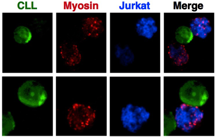
CLL cells associate with myosin exposed apoptotic cells (MEACs). Confocal microscopy of representative CLL cells (green) associated with MEACs (induced in a human Jurkat T cell line (blue)) with exposed Myosin on its cell surface (red) shown as separate images for each color and then merged together (Merge). (Reference Cui et al. Leukemia 2016)
Our long-term goal is to understand the properties of antibodies produced by CLL cells and the nature of the molecular and cellular targets to which these antibodies bind. This understanding may also further define the cell of origin and CLL. This new knowledge will ultimately lead to developing better therapeutic strategies to eliminate this disease.