Resting State fMRI in Adult Macaques: Validating an Approach
What is resting state fMRI? What does it measure, and what constrains it?
 In humans, correlations between spontaneous activity in specific nodes of the CNS, measured via resting state functional connectivity MRI (rs-fMRI), are increasingly being tested as predictors of disease states or disease endotypes in humans. Subjects are scanned while ‘at rest’, i.e. awake but not performing a task. The meaning of correlated spontaneous activity under these circumstances, and its relationship to true anatomic connectivity, remains to be validated. For example, it is uncertain whether aberrant functional ‘connectivity’ derives from direct neuron-to-neuron communication deficits, indirect circuits, or artifact. Moreover, it is not clear whether internal ‘states’ of individuals (stress, pain, anticipation) drive resting state patterns. rs-fMRI studies in nonhuman primates are valuable in two ways.
First, a known limitation of rs-fMRI connectivity analysis is that the physical substrates (i.e. neural projections) underlying the proposed connections are often not known and cannot be verified across and within human subjects. Second, current connectivity analysis largely relies on Pearson correlation as a metric for connectivity, a method that is unable to convey information about the direction of connections and thereby limits our understanding of information flow in the brain. Overcoming these barriers requires the use of more intricate rs-fMRI analysis techniques, such as Granger Causality, as well as an animal model in which proposed connections can be verified using tract tracing.
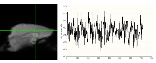
This project involves protocol development to validate resting state functional connectivity (rs-fMRI) paradigms using anesthetized nonhuman primates (Old world monkeys). We are collecting data on the Siemens Prisma 3T MRI scanner, in stereotaxically placed animals under low level anesthesia (Vincent et al 2007). We subsequently place precise injections of neuronal tracers in selected brain regions, and in order to map connections at the microscopic levels. These maps in turn will be re-aligned onto MRIs in order to precisely localize seed placements, and make hypotheses about strength and directionality of rs-fMRI outcomes.