Integrating Social Networks Through the Amygdala
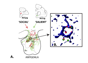
Figure 1. A. Map of anterogradely labeled terminals from the ACCsg (green) and ACCpg (red), representing ‘salience’ and ‘social monitoring’ nodes of the prefrontal cortex respectively, in the amygdala. Overlap of terminals occurs in specific amygdala subregions, where higher power confocal imaging (B.) was then conducted to examine whether terminals predominantly contact the same, or different subpopulations of projection neurons labeled with CAMIIa (blue, marker of projection neurons). Axon contacts from the ACCpg (red) and ACCsg (green) frequently contact the same neuron and lie in close apposition, suggesting tight coordination of cell excitability.
The salience network is an interconnected set of brain ‘hubs’ that active together during arousing emotional states. In contrast, the ‘social’ brain hubs are considered more ‘cognitive’ and are active when subjects observe emotion in others. These networks appear to operate separately to tag different tasks at the cortical level in functional imaging studies. However, social information has the highest level of salience for humans. Psychiatric syndromes are frequently characterized by miscoding the intensity, relevance, valence, or shifts of social cues—in short, their salience. Integration of these paths must therefore occur.
We are examining the connectional details of how the ‘social brain’ nodes interact with ‘salience detection’ networks through afferents to the amygdala. The amygdala is a key site for emotional coding of various cues, including faces. Tract tracing studies in nonhuman primate enable more accurate interpretation of neuroimaging results in humans, and are therefore a critical bridge for understanding details of primate brain structure on a cellular level. We recently found that the ‘salience detection’ node of the prefrontal cortex (the subgenual anterior cingulate, sgACC) which monitors internal physiologic states to ‘mark’ salient cues, and the ‘social monitoring’ network (the pregrenual anterior cingulate, pgACC) which detects and interprets the meaning and value of others’ actions, send overlapping inputs to specific regions of the primate amygdala. Interestingly, the salience node afferents have a much broader input to the amygdala, consistent with a broader regulatory role. Yet, ‘social’ brain node inputs consistently overlap ‘salience’ network afferents. This indicates that in the amygdala, social monitoring information is always coded in the context of ‘salience’ information. We are currently working to understand how the 2 networks influence cellular populations. Do intermingled cells receive one set of inputs or the other? Or do these two ‘separate’ nodes converge on the same neurons, and to what extent?