Role of Neuroinflammation in Methamphetamine Induced Neurotoxicity
Project Collaborator:
Dr. Lisa Opanashuk

Local striatal neuroinflammation (MHC II
staining) following FIV-cre injection in IL-1β XAT mice.
Long-term methamphetamine abuse is associated with significant impairment of brain function, likely arising from degeneration of specific neuronal populations, including dopaminergic cells innervating the striatum as can be modeled in rodents exposed to methamphetamine. We are exploring the hypothesis that methamphetamine associated neuroinflammation contributes to the neurotoxic insult and may be an important source of oxidative stress believed to play a role in tissue damage. With funding from the National Institute of Drug Abuse we are investigating the specific roles of IL-1 and microglia mediated oxidative injury in striatal neurodegeneration following methamphetamine administration in mice.
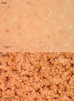
Striatal astrocyte activation (GFAP staining) 7
days after acute methamphetamine exposure.
The studies have special implications for individuals with HIV who also abuse methamphetamine since HIV infection can lead to a devastating neurological impairment known as AIDS dementia that includes a prominent neuroinflammatory component. Indeed, while our studies do not directly address the synergy between HIV infection and methamphetamine, our project includes experiments exploring the effects of drug toxicity in the setting of pre-existing neuroinflammation. By identifying processes and specific mediators involved in neurotoxicity, these studies may provide opportunities for preventing some of the damage occurring with drug abuse.