URMC / Labs / Robert Lab / Projects / Evolution of Host/Viral Pathogen Interaction
Evolution of Host/Viral Pathogen Interaction
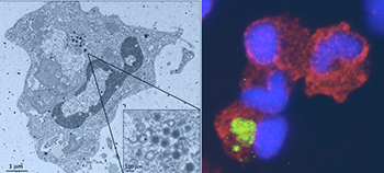
(Left) Electron microscopy view of a Xenopus peritoneal
macrophage infected by FV3. (Right) Persistent FV3 infection in
peritoneal macrophages detected by Fluorescence microscopy.
Green: FV3 (anti-53R); Red: macrophage (anti-HAM56); Blue: nuclei
(Hoechst-33258).
1) Immune responses to emerging Ranaviral diseases: This research area concerns basic comparative and applied studies of viral pathogenesis and immunity in amphibians caused by Poxvirus-like Ranaviruses such as Frog virus 3 (FV3). Because of the threat of emerging wildlife viral diseases on global biodiversity, fundamental research on comparative viral immunity has become crucial. We have established Xenopusas an important experimental model to study the host defense and the pathogenesis of ranavirus infection, and evaluate the contribution of immunocompromised animals in the dissemination of the diseases.
In collaboration with Brian Ward (URMC), we have developed an efficient and reliable method to knockout (KO) putative virulence genes by site-specific integration of a selectable fluorescent marker into the viral genome. Susceptible Xenopus larvae provide an ideal model to evaluate the impact of KOs in vivo on virus load, host mortality and the induction of pro-inflammatory genes.
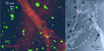
Neo-vascularization is induced in 15/0 semi-solid tumor grafts.
2) Role of macrophages in ranavirus infection: We have shown that during ranavirus infection macrophages play a critical role in orchestrating antiviral and inflammatory responses as well as antigen-presenting cells, promoting CD8 T cell responses. However, despite efficient viral clearance, macrophages can also harbor quiescent, transcriptionally inactive, virus within otherwise healthy asymptomatic frogs. In collaboration with Brian Ward (URMC), we have developed an efficient and reliable method to knockout (KO) putative virulence genes by site-specific integration of a selectable fluorescent marker into the viral genome. Susceptible Xenopus larvae provide an ideal model to evaluate the impact of KOs in vivo on virus load, host mortality and the induction of pro-inflammatory genes.
We have developed reagents (specific antibodies, recombinant cytokines including type I IFN, CSF-1 and IL-34) and transgenic strains (with cells of the myeloid lineage or macrophage expressing the GFP reporter) to characterize the involvement of macrophages in tadpoles and adults during ranavirus infection. We also found that we can reactivate persistent but asymptomatic FV3 infection by providing pro-inflammatory signals recruiting macrophages in the peritoneal cavity.
3) FV3/Xenopus tadpoles model system for studying blood brain barrier integrity. In collaboration with Sanjay Maggirwar, we have investigated whether inflammation caused by FV3 in tadpole affect the blood brain barrier (BBB) integrity. We found indeed that FV3 infection results in BBB defects, leukocyte infiltration, increase TNF and Il-1 gene expression and viral dissemination into neural tissues of tadpoles but not adult Xenopus. We propose to take advantage of the transparency and accessibility to experimentation of tadpoles, to develop a new platform for investigating effects of viral infection and inflammation on BBB integrity.
« back to all projects