How are sodium channels clustered at nodes of Ranvier?
Model for node of Ranvier formation
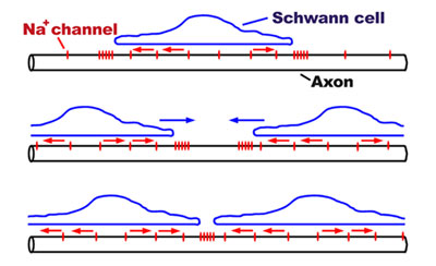
Our laboratory has developed an hypothesis for node of Ranvier formation and we continue to test these ideas. Prior to myelination (and following demyelination) sodium channels are present throughout the axon membrane at low density. A former graduate student, Jin Wu, characterized these and other axonal channels with patch clamp measurements. When glial cells (Schwann cells in the peripheral nervous system) adhere to axons and begin myelination, sodium channels rapidly cluster adjacent to the tips of the glial processes that are extended during early stages of ensheathment. One idea, illustrated in the top sketch, is that the axonal sodium channels are excluded from regions of close association with Schwann cells, and they then diffuse laterally and collect just outside the process tip.
As Schwann cells grow longitudinally, the channels appear to move with them, since the distance across the glial cell grows, and the clusters remain always just past their edges (middle sketch). Sodium channel clusters associated with neighboring Schwann cells thus move closer and closer together, ultimately fusing with a neighboring cluster to form a node of Ranvier (bottom sketch). This model was developed by former postdoctoral fellow Sanja Novakovic, and by recent graduates Elda Tzoumaka, Ian Vabnick, and Matthew Rasband. While we have much evidence for the cellular events depicted in this model (see references below), we seek to identify the molecular mechanisms responsible for this neuron-glial communication. Graduate students Katie Kazarinova-Noyes and Andrew Custer have recently identified 2 candidates, both membrane proteins in the immunoglobin superfamily, and we are testing their involvement in these events.