Measurements of cerebrovascular reactivity using BOLD fMRI and ASL
Measurements of cerebrovascular reactivity using BOLD fMRI and ASL
Cerebrovascular reactivity (CVR), the mechanism that reflects the compensatory change in blood flow in response to a vasoactive stimulus, is an important marker of vascular health. Alterations of CVR have been associated with vascular pathophysiology, such as vasculitis, carotid artery disease, hypertension, arteriovenous malformations, traumatic brain injury, moyamoya vasculopathy, seizures, and cognitive decline. Thus its measurement is critical in determining outcomes and treatment options. The goal of the project is to evaluate non-invasive ways of measuring cerebrovascular reactivity using BOLD MRI techniques.
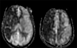
Figure 1: Cerebral Blood Flow map obtained with ASL in a patient with right ICA/MCA stenosis.

Figure 2: Cerebral Vascular Reactivity map obtained with BOLD fMRI in the same patient.