Characterization of Trabecular Bone Micro-architecture for Bone Strength Estimation
Characterization of Trabecular Bone Micro-architecture for Bone Strength Estimation
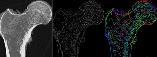
(LEFT) MDCT image of the proximal femur (MIDDLE) same image but pixel values are now
indicative of the magnitude of local structural anisotropy (RIGHT) pixel values are now indicative of
the direction of local structural anisotropy. We investigate the hypothesis that a characterization of
trabecular bone structure that incorporates such bone anisotropy can improve biomechanical
strength prediction in the proximal femur.
We are investigating the role that texture features can play in characterizing the trabecular bone micro-architecture of the proximal femur or tibia, and spinal vertebrae, as visualized through multi-detector CT (MDCT) or MRI. We are particularly interested in using such texture features, indicative of local topology or geometry of underlying bone tissue structures, as quantitative measures for predicting bone strength. Such measures could serve as imaging bio-markers for predicting the likelihood of osteoporotic hip fractures, which can seriously compromise the health and quality of life for patients suffering from osteoporosis.
Where other researchers have investigated the correlation between bone strength and different features characterizing the trabecular bone, we are specifically interested in using such features (extracted in a non-invasive manner) in conjunction with machine learning techniques for predicting the strength of the femur. Our long term goal is to establish a CADx framework for automated bone strength prediction through non-invasive analysis of the trabecular bone regions of patients with osteoporosis. We also pursue approaches that characterize the anisotropy of the trabecular bone micro-architecture. The distribution of trabecular bone has been shown to be highly anisotropic (as seen on image shown to the right) and texture features that take this into account could potentially serve as more powerful predictors of bone strength over conventionally used features related to bone mineral density (BMD).
Journal Publications
- C.-C. Yang, M.B. Nagarajan, M.B. Huber, J. Carballido-Gamio, J.S. Bauer, T. Baum, F. Eckstein, E. Lochmüller, T.M. Link, and A. Wismüller, "Improving bone strength prediction in human proximal femur specimens through geometrical characterization of trabecular bone microarchitecture and support vector regression," Journal of Electronic Imaging 23(1):013013 (2014).
- M.B. Huber, S.L. Lancianese, M.B. Nagarajan, I.Z. Ikpot, A.L. Lerner and A. Wismüller, "Prediction of Biomechanical Properties of Trabecular Bone in MR Images with Geometric Features and Support Vector Regression," IEEE Transactions on Biomedical Engineering, 58(6):1820-1826, (2011).
Conference Publications
- C.-C. Yang, M.B. Nagarajan, M.B. Huber, J. Carballido-Gamio, J.S. Bauer, T.H. Baum, F. Eckstein, E.-M. Lochmüller, T.M. Link, and A. Wismüller, "Predicting the biomechanical strength of proximal femur specimens with Minkowski functionals and support vector regression," Proceedings of SPIE Medical Imaging 9038:101-108 (2014).
- C-C. Yang, M.B. Nagarajan, M.B. Huber, J. Carballido-Gamio, J.S. Bauer, T. Baum, F. Eckstein, E. Lochmüller, S. Majumdar, T.M. Link, A. Wismüller, "Predicting the Biomechanical Strength of Proximal Femur Specimens through High Dimensional Geometric Features and Support Vector Regression," Proceedings of SPIE Medical Imaging 8672:1K1-1K8 (2013).
- M.B. Huber, C. Yang, J. Carballido-Gamio, J.S. Bauer, T. Baum, M.B. Nagarajan, F. Eckstein, E. Lochmüller, S. Majumdar, T.M. Link, and A. Wismüller, "Predicting the biomechanical strength of proximal femur specimens with bone mineral density features and support vector regression," Proceedings of SPIE Medical Imaging 8315:1W1-1W7, (2012).
- M.B. Huber, S.L. Lancianese, I.Z. Ikpot, M.B. Nagarajan, A.L. Lerner and A. Wismüller, "Prediction of biomechanical trabecular bone properties with geometric features using MR imaging," Proceedings of SPIE Medical Imaging 7626:1O1-1O8, (2010).