Pituitary Tumors (Benign)
Make Appointments & Get Care
What are Pituitary Tumors (Benign)?
The pituitary gland is the “master gland,” coordinating multiple hormone systems. Pituitary tumors develop in about 10% of the population, and almost all are benign (non-cancerous).
But these benign tumors, called adenomas, can cause the pituitary gland to produce too low or too high levels of hormones. Types include:
- Non-functioning pituitary adenoma. These tumors have a number of different names, but they all mean the same thing: the tumor is not causing excess production of a hormone but instead is an abnormal, benign tissue growth that causes problems by taking up space. When the growth is greater than 10 mm in diameter, it can compress the pituitary gland, leading to insufficient pituitary functioning.
- Prolactinoma. This common benign tumor causes too much of a normal hormone, prolactin, which is important for the normal production of breast milk in women who are nursing. Otherwise, it’s present at low levels in both men and non-pregnant or non-nursing women.
- Acromegaly and gigantism. In acromegaly, a benign tumor produces too much growth hormone in an adult. Gigantism is a similar condition but is specific to children who are still able to grow abnormally in response to the abnormally high amount of growth hormone produced by the tumor. When these tumors occur in adults, they usually appear in middle age. Acromegaly is associated with hypertension, diabetes mellitus, and cardiovascular disease.
- Cushing’s disease. Cushing syndromes result from long-term and excessive exposure to corticosteroids, causing physical and emotional symptoms. Abnormalities in the hypothalamus, pituitary gland, and/or adrenal glands can cause the balance of cortisol to be disturbed, leading to Cushing’s syndrome.
By the Numbers
200+
New patients treated yearly
Largest referral program in New York
Schedule an appointment with a UR Medicine provider.
Call (585) 275-2901UR Medicine's Treatments for Pituitary Tumors (Benign)
The UR Medicine Pituitary Program provides the Rochester metropolitan area and surrounding region's most advanced and comprehensive care to patients with pituitary and hypothalamic disorders. As part of a leading academic medical center, we bring together a team of specialists from several areas of medicine to care for our patients.
Our experts in neurosurgery, endocrinology, radiation oncology, and neuro-ophthalmology help patients and their families navigate the complex issues surrounding disorders of the pituitary gland and hypothalamus. We use the newest technologies and treatments available in the world, and we deliver care with compassion and an understanding of the many complexities involved in treating patients with these disorders.
Patient Stories
Police Chief Receives life-saving surgery
Diagnosed with Cushing's disease, local police chief found hope at UR Medicine.
Surgery Offers Relief for One Woman's Unsolvable Symptoms
Providers at UR Medicine offered Amy relief with a diagnosis and surgery for prolactinoma, a small, benign pituitary tumor, which had caused seemingly unsolvable symptoms.
What Sets Us Apart?
- Studies have shown that hospitals treating more than 25 pituitary tumors per year had significantly better outcomes*. The UR Medicine Pituitary Program routinely treats over 200 new patients a year, making us one of the most experienced centers for the diagnosis and treatment of pituitary tumors in the Rochester metropolitan area and surrounding region.
- Our program is the largest referral program in New York and the only one within 300 miles of Rochester that provides true multidisciplinary care to patients with pituitary disorders.
- Our team evaluates all disorders affecting the function of the pituitary gland and hypothalamus and coordinates all aspects of care.
- The program is able to offer simultaneous neurosurgical and endocrinologic evaluation for patients. Patients also have access to a complete team of specialists in radiation oncology, neuro-ophthalmology, neurology, reproductive medicine, and genetic counseling.
- Our team offers patients and family members the caring support they need throughout their treatment and actively supports the Pituitary Network Association.
*Chen, C., Hu, Y., Lyu, L. et al. Incidence, demographics, and survival of patients with primary pituitary tumors: a SEER database study in 2004–2016. Sci Rep 11, 15155 (2021). https://doi.org/10.1038/s41598-021-94658-8
Locations
View All LocationsWe serve you in the Rochester metropolitan area and surrounding region.
View All Locations4 locations
2180 South Clinton Avenue
Rochester, NY 14618
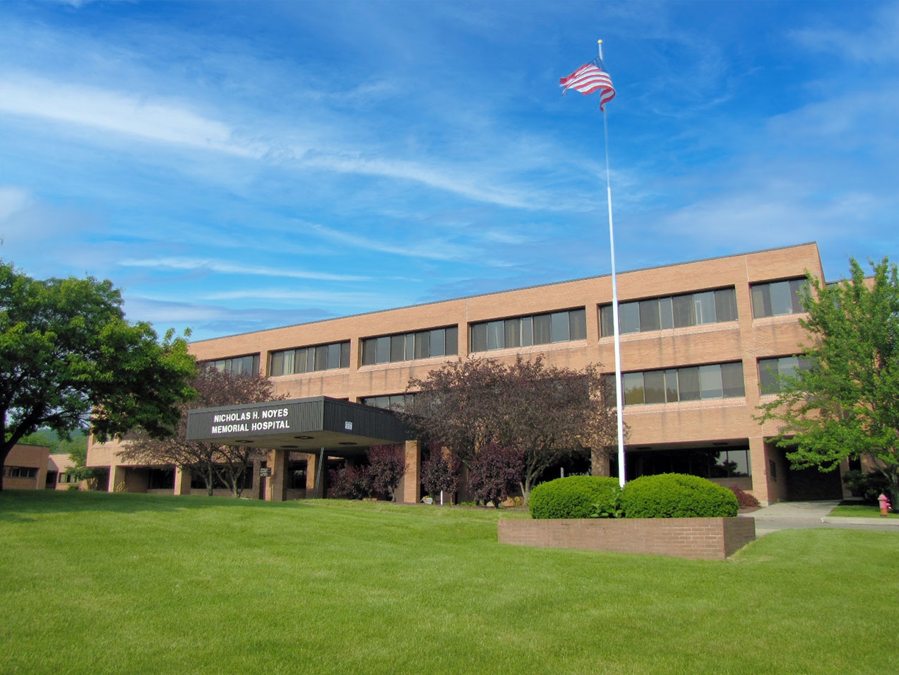
Noyes Memorial Hospital
111 Clara Barton Street, Suite 178A
Dansville, NY 14437
1340 Washington St, Suite 3
Watertown, NY 13061
