Pathology@URMC Blog

Not Just Cut and Dry: A Day in the Life of a Histologist
Behind many patient diagnoses is a process that starts in the operating room and ends under a microscope.
UR Medicine Labs has a team of histotechnicians (HT) and histotechnologists (HLT), often referred to as "histotechs," who work around the clock to complete the steps leading up to patient diagnoses for cancer and other diseases.
Histotechs are responsible for taking human tissue to prep, cut, and stain before it's examined by a pathologist. We recognize this important work in honor of Histotechnology Professionals' Day on Thursday, March 10.
"I think most people have never heard of histology," said histotech Wally Pryjmak, who has worked at UR Medicine Labs for two years. He didn't even know what a histologist was when he became a certified medical lab technician. Nevertheless, Pryjmak wants patients to understand the link between his work and their treatment. As he says, "We create the slides your diagnosis is based on, so without us it would be a lot harder to get diagnosed."
The tissue prep process starts after a patient has surgery to remove tissue from the body, often in a biopsy. This tissue is brought to the Surgical Pathology area (located on the ground floor of SMH) and is most often examined first by a pathologist's assistant (PA) who dissects the tissue at a grossing station.
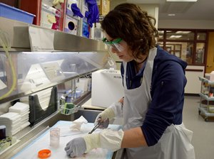
The tissue sample then goes into the hands of a histologist who processes it through a series of alcohols to remove any water, clears tissue in xylene and infiltrates it in paraffin wax. The tissue is then embedded into a paraffin block and the histologist cuts it into paper-thin slices.
Wax slices of tissue then go into a warm water bath to stretch them out and prevent wrinkles before carefully placing the tissue onto a glass microscope slide. The histologist gently scoops the section of tissue onto the slide to dry. He or she then treats the slide with a basic stain (hematoxylin and eosin) to make the sample is easy to see under the lens.
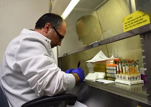
There are up to 75 different stains that reveal certain tissue elements, fungus, or bacteria. A pathologist can request any combination of stains be added before viewing the slide close-up.
In this way, the histologist is part artist and part chef: they must follow a "recipe" of stains to yield a proper reading. Even a small misstep can compromise the diagnosis. Once the slide is treated with the proper stains, it goes to a pathologist who examines it and issues the diagnosis.
The work of a histologist requires careful attention to detail and physical stamina. On average, the histology team at Strong prepares between 800 and 1,200 slides each day.
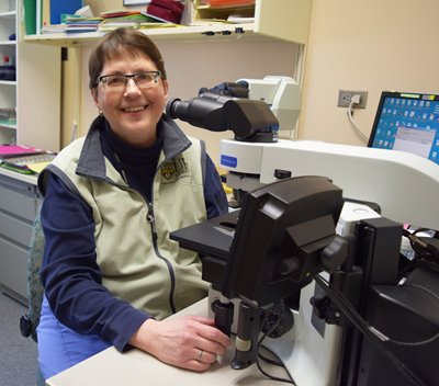
Diana Scott is the supervisor for histopathology at Strong. To her, the work of a histologist is like an art form. And while patients don't see or speak with the lab team helping behind the scenes, the connection is still very meaningful.
"We care about the production side of things and the quality of the slides," said Scott. "We treat every specimen like it belongs to one of our family members waiting for their test results to come back."
In photos:
(Top) Histologist Lisa Torregrossa embeds a tissue sample into paraffin wax. Once the wax hardens, the block can be sliced for examination. (Top right) Elizabeth Pilon uses a microtome instrument to slice a piece of human breast tissue that has been embedded in wax.
(Above right) There are more than 75 special stains used to dye tissues so they can be viewed clearly under a microscope. The stains highlight specific tissue components (nuclei, muscle, etc.) or microorganisms (bacteria, fungus) and a combination of stains is often necessary.
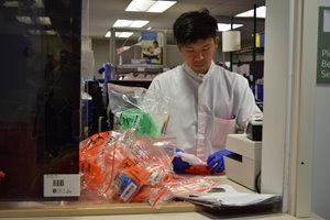
(Right) Tissue blocks, or cassettes, are preserved and filed for at least 20 years after a diagnosis is given.
Bethany Bushen | 3/7/2016