Patient with Recurrent Microhematuria
Patient with Recurrent Microhematuria
Hangchuan Shi, MD, PhD, Veronica Ulici, MD, PhD, Hiroshi Miyamoto, MD, PhD
Past Medical History
A 58-year-old male with a history of BPH and prostate adenocarcinoma (Gleason 3 + 4), treated with high dose rate brachytherapy twice.
Present History
The patient presented with recurrent microhematuria. Cystoscopy revealed diffuse cystic nodules, each measuring approximately 2 mm, scattered throughout the bladder.
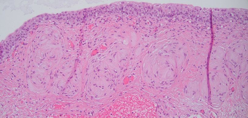
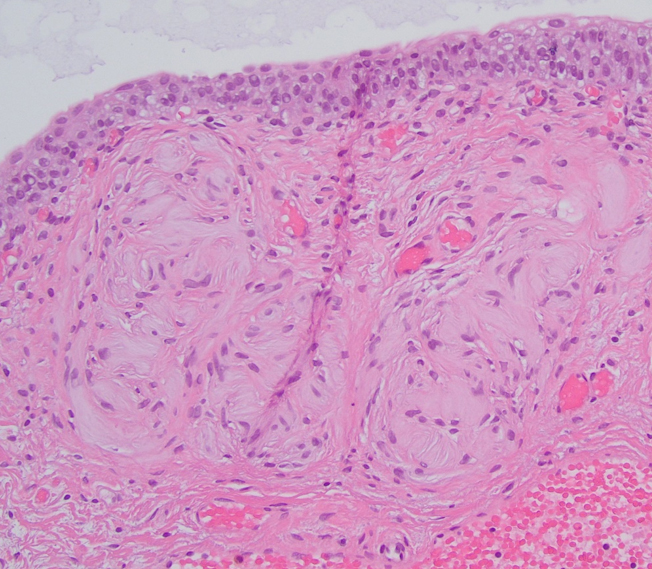
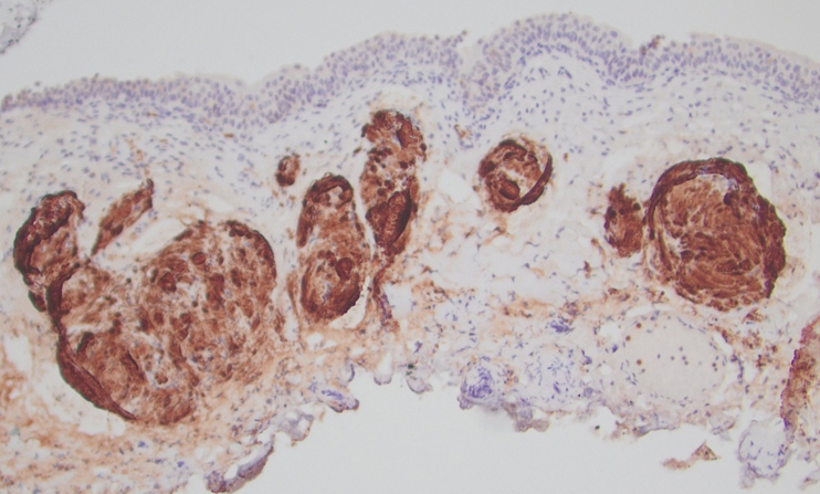
Pathology
Transurethral resection of the lesion revealed a normal urothelial mucosal lining. Within the submucosa, multiple pink, round structures of varying sizes were identified. These structures demonstrated concentric lamellar layers of eosinophilic material, surrounded by Schwannian cells (images 1,2,3). Immunohistochemical stains showed that the cells were positive for S100 (image 4), SOX10, and CD34.