Young Female with Chronic Cutaneous Ulcers and Sinonasal Lesion
Young Female with Chronic Cutaneous Ulcers and Sinonasal Lesion
Anna Savin, MD; Richard Burack, MD, PhD; Kathleen Mannava, MD
Clinical History
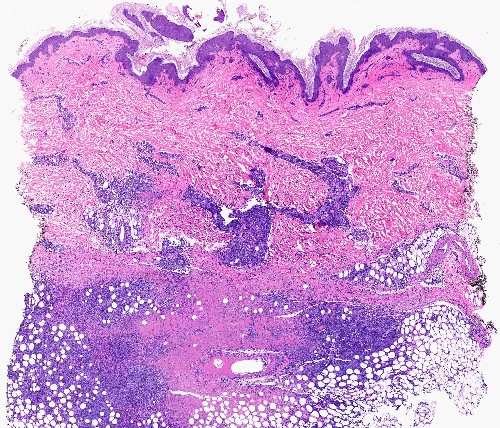
A young woman with no significant past medical history presented with chronic non-healing ulcers involving the foot and ankle. Skin biopsies demonstrated dense angiodestructive inflammation and coagulative necrosis, supporting the clinical impression of medium-vessel vasculitis (Figure 1). Despite treatment, the lesions persisted. Several months later, the patient developed fever, nasal congestion and purulent drainage, requiring hospitalization. The patient underwent endoscopic sinus surgery with debridement.
Microscopic Findings
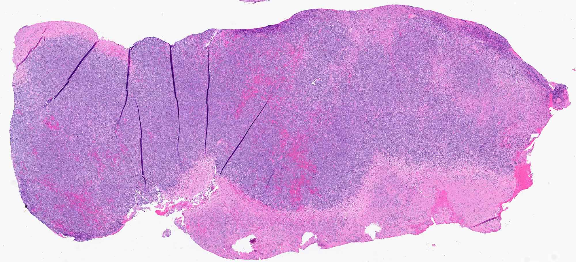
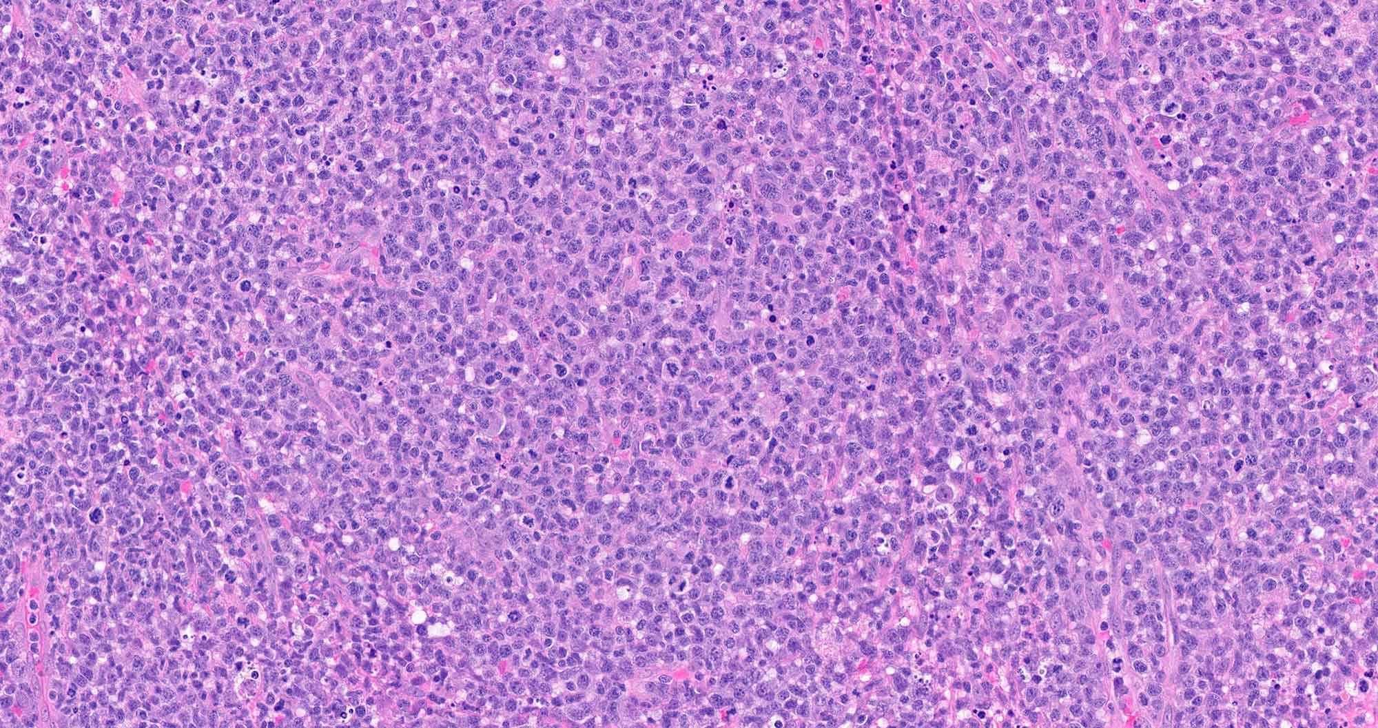
Histologic examination of the sinonasal tissue demonstrates an atypical lymphoid infiltrate associated with extensive coagulative necrosis and ulceration (Figure 2). The atypical cells are intermediate in size with irregular nuclear contours, pale cytoplasm, frequent mitotic figures, and abundant apoptotic debris (Figure 3). An angiocentric and angiodestructive growth pattern is noted.
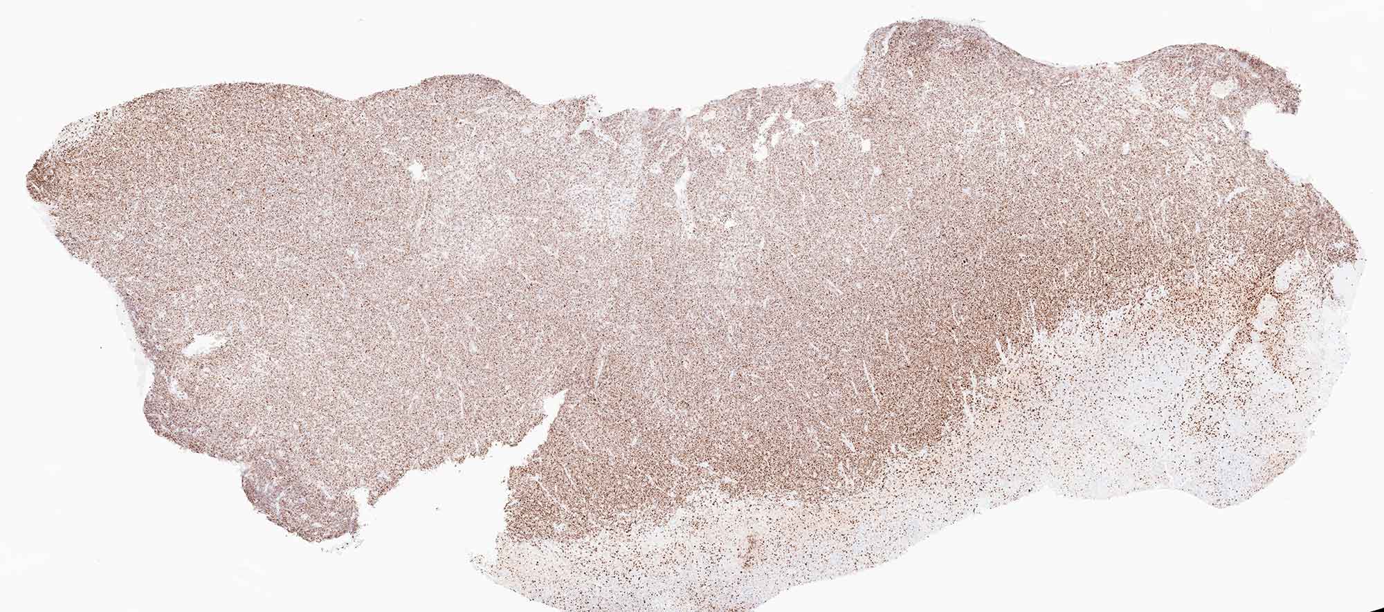
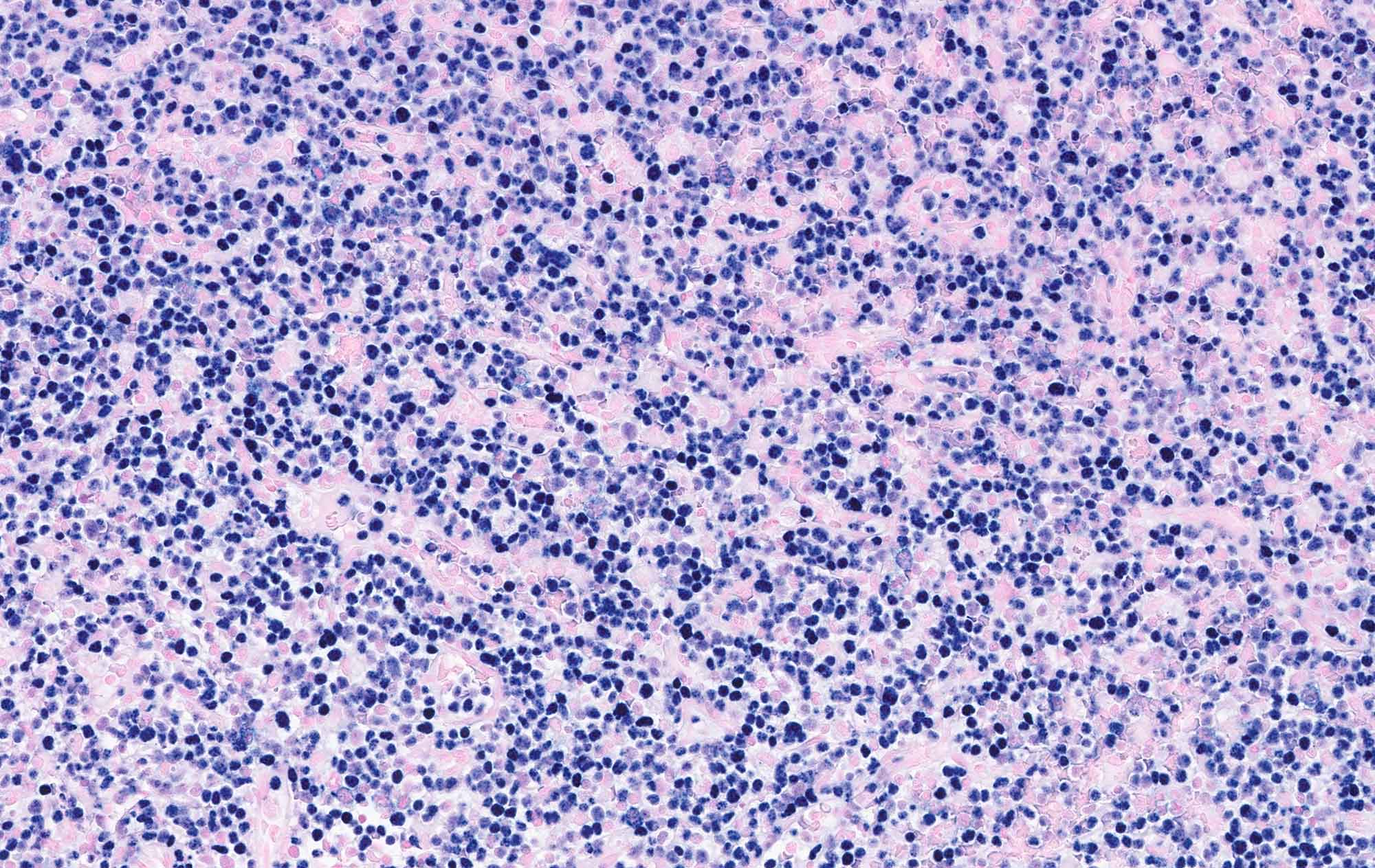
Immunohistochemical studies demonstrated expression of CD56, TIA-1, granzyme B (Figure 4), and CD3. EBER in situ hybridization showed diffuse positivity within the neoplastic cells (Figure 5). The Ki-67 proliferation index approached 95%.