Middle Aged Male with Hilar Lesion
Middle Aged Male with Hilar Lesion
Tausif Ahmed Siddiqui, B.S.; Moises J Velez, M.D.
Clinical History
A 65-year-old man with a history of hypertension, hyperlipidemia, gastroesophageal reflux disease, gout, and obstructive sleep apnea was incidentally found to have a right hilar lesion on a cardiac CT calcium score study. A subsequent chest CT demonstrated a 1.7 cm right infrahilar lesion, initially favored to represent an enlarged hilar lymph node. Follow-up imaging showed interval enlargement to 3.3 × 1.9 cm. Endobronchial ultrasound-guided fine needle aspiration demonstrated a low-grade neuroendocrine neoplasm positive for synaptophysin, chromogranin, and INSM1, with weak focal TTF-1 expression and negative Napsin A and p40. The Ki-67 index was approximately 5%. DOTATATE PET/CT demonstrated mild uptake in the right hilar lesion without DOTATATE-avid regional lymphadenopathy.
The patient underwent robotic-assisted right middle and lower bilobectomy with mediastinal lymph node dissection. Gross examination revealed a 2.5 × 2.0 × 1.9 cm ill-defined hilar mass with a gray white to black cut surface and focal hemorrhage and yellow discoloration. The tumor invaded the bronchial wall and hilar vessels and was 0.3 cm from the closest hilar vascular margin.
Microscopic Findings
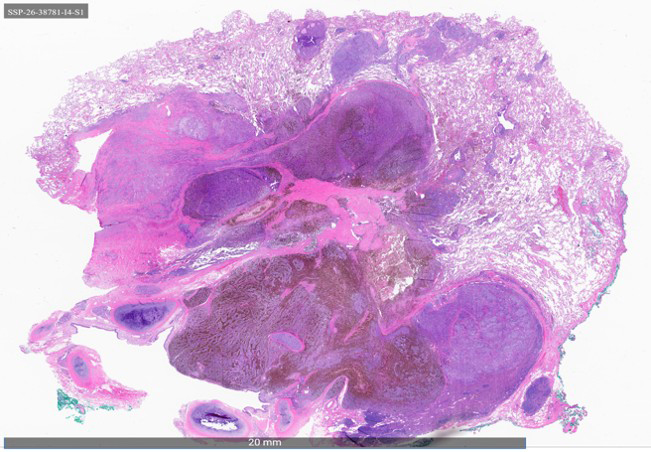
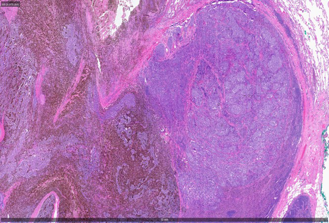
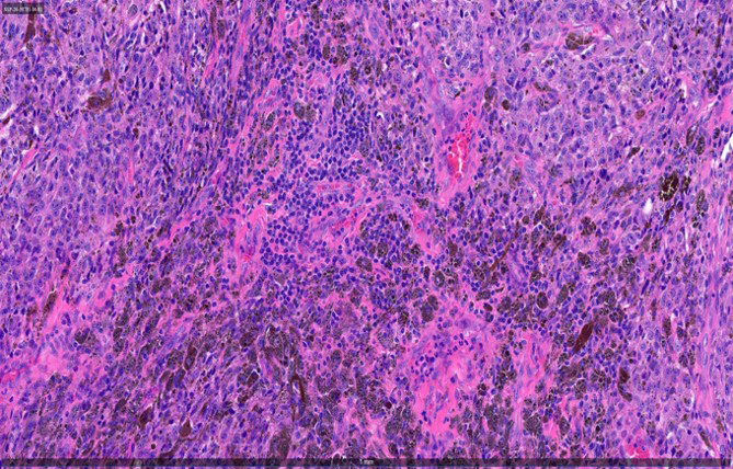
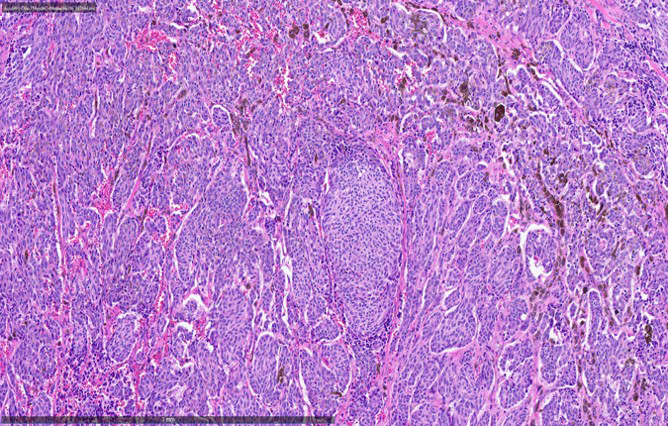
Histologic sections demonstrated an epithelioid and spindle cell neoplasm with an organoid growth pattern (Figures 1A-D). The tumor showed abundant brown-black intracytoplasmic melanin pigment, readily apparent on H&E and highlighted by the melanin stain (Figure 1E). At higher magnification, the tumor was composed of epithelioid and spindle cells with abundant intracytoplasmic melanin pigment and focal pseudorosette-like architecture (Figures 1C-D).
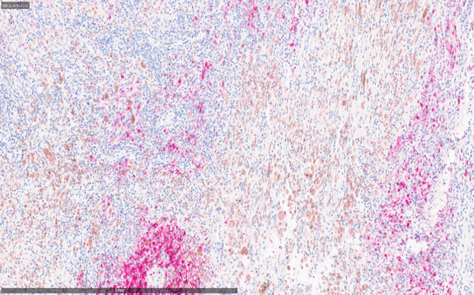
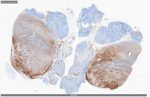
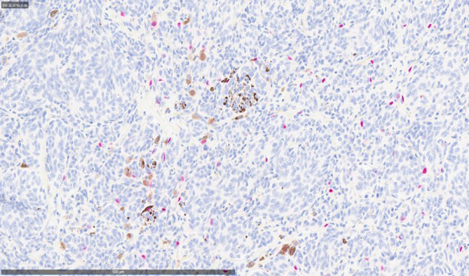
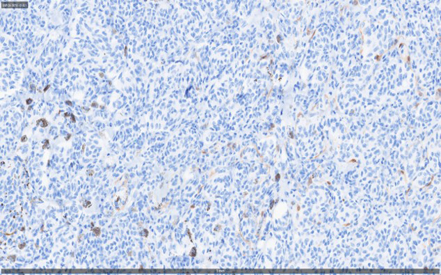
Immunohistochemistry demonstrated diffuse pancytokeratin (PanCK) expression in the neoplastic cells, supporting epithelial differentiation (Figure 1F). SOX10 highlighted a subset of neoplastic cells, supporting focal melanocytic differentiation (Figure 1G). S100 highlighted a population of sustentacular cells surrounding the tumor nests (Figure 1H). S100 and SOX10 are both used as melanocytic markers and may also highlight sustentacular cells; therefore, the distribution of staining is important for interpretation. The presence of sustentacular cells was consistent with the characteristic architecture of the tumor, while SOX10 expression in a subset of neoplastic cells supported divergent melanocytic differentiation.