Blood Cell Imaging
The eye may offer a noninvasive alternative to drawing blood
Most people cringe when their blood is drawn by a needle and syringe for testing. They bear it anyway because of the "diagnostic potential in a single drop of blood," says Jesse Schallek, Assistant Professor of Ophthalmology. But what if, instead of drawing the blood, we could instead "leave the blood cells inside the body where they naturally reside, and study them inside living tissue?" Schallek asked at the recent Falling Walls competition.
What if at least some blood tests could be as noninvasive as an eye exam?
The neural cells that line the back of our eyes are supported by a dense network of capillaries that circulate blood to deliver nourishment and remove waste.
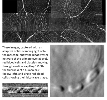
Schallek's lab, in collaboration with colleagues at the Flaum Eye Institute, the Center for Visual Science, and The Institute of Optics, uses a specialized camera called an Adaptive Optics Scanning Light Ophthalmoscope (AOSLO) — which can correct for small imperfections of the optics of the eye — to obtain images of retinal blood vessels that are ten-times thinner than a human hair. Videos can even capture the movement of single blood cells flowing within this network.
"We have learned interesting tricks from using adaptive optics," Schallek said. "We can take snapshots of single red blood cells, showing their concave shape; we can combine adaptive optics with high temporal resolution to see a virtual fire hose of information from these tiny capillaries."
His lab is also evaluating different biomarkers — such as the interaction properties of light, including its spectra and light scatter — to evaluate cell size and differentiate which types of blood cells are present. This information may eventually provide a complete blood count (CBC) that examines ratios of red blood cells, white blood cells and platelets — all within the living body.
Imaging flow cytometer in the living retinal vasculature
A current graduate student in Schallek’s lab, Jin Huh, is exploring the potential for using AOSLO to enable in vivo flow cytometry of fluorescently-tagged white blood cells. Typical blood analysis involves sending a blood sample to a dedicated processing facility. In our lab AOSLO enables us to visualize fluorescently-labeled blood cells inside retinal blood vessels directly. This method may one day allow humans to forego a blood draw as we are now able to assess the systemic health of the subject through the living eye.

Schallek's work also uses new approaches to measure blood cell speed by tracking the velocity of cells over time. This information is of critical importance to identify vascular dysfunction of the eye, a leading cause of blindness in the developed world.
Schallek and his team are working collaboratively with the Departments of Ophthalmology and of Biomedical Engineering, the Institute of Optics, and the Center for Visual Science to refine this technique and begin measurements in human patients. Because the AOSLO approach is non-invasive and uses safe levels of light for measurement, Schallek's team is directly bringing their design from concept to deployment in a bench-to-bedside translational project this summer.
There are many targets to evaluate in patients. Among the earliest Schallek will identify are blood cells in patients with leukemia, sickle-cell anemia and those on blood thinners, all of which can change the ratios and properties of blood cells in the living body.