Mammogram
Make Appointments & Get Care
What Is a Mammogram?
A mammogram is a specialized X-ray used to examine breast tissue. It plays a key role in both the early detection and diagnosis of breast conditions, including breast cancer. Mammograms can be performed as a screening tool—even when there are no noticeable symptoms—or as a diagnostic test to evaluate changes such as breast pain, lumps, or nipple discharge.
The procedure uses a low dose of radiation to create detailed images of the breast, helping radiologists identify abnormalities that may not be felt during a physical exam. Regular screening mammograms are recommended for many women beginning at age 40, though individual risk factors may lead to different recommendations.
During a mammogram, your breasts are squeezed by the equipment for a few seconds, which may cause mild pain. This pressure keeps the radiation low and helps take the best picture of your breast.
The Tyrer-Cuzick Risk Assessment Calculator, a nationally accepted standard based on science, helps to measure women’s breast cancer risk factors based on family history, and helps women to better understand their breast cancer risk factors. It allows health professionals to estimate a woman’s risk of developing invasive breast cancer over the next five years and up to age 90.
*This tool can’t accurately estimate breast cancer risk for women:
- Carrying a breast-cancer-producing mutation in BRCA1 or BRCA2
- With a previous history of invasive or in-situ breast cancer
- Lobular cancer in- situ (LCIS)
- Ductal cancer in-situ (DCIS)
- In certain other subgroups
Schedule your mammogram online.
Important: Online appointments are for screenings only. We will contact you within 3-5 business days to schedule your appointment.
What Conditions Does A Mammogram Show?
A mammogram can show the following conditions:
Calcifications are tiny mineral deposits in the breast tissue. There are 2 types of calcifications:
- Macrocalcifications — Larger calcium deposits that often mean worsening changes in the breast.
- Microcalcifications — Tiny (less than 1/50 of an inch) specks of calcium.
Masses, sometimes called a breast lump, may happen with or without calcifications. Masses may be caused by:
- A cyst — A noncancer (benign) collection of fluid in the breast
- Benign breast conditions
- Breast cancer
Screening vs. Diagnostic Mammogram: What's the Difference?
A screening mammogram is a routine test for people with no signs or symptoms of breast health conditions. It is used to check for early signs of breast cancer, often before it can be felt.
A diagnostic mammogram is used when there’s a known concern—like a lump, pain, or unusual changes on a screening mammogram. It involves more detailed images to help get a closer look at the area.
Who Should Get A Screening Mammogram?
Different health experts have different recommendations for people who have no symptoms of breast cancer:
- The U.S. Preventive Services Task Force recommends screening mammograms every 2 years for women ages 40 through 74. This applies to people at average risk of breast cancer.
- The American Cancer Society recommends screening starting at 40 for those of average risk. Mammograms should be done every year for everyone ages 45 to 54. Then you may switch to mammograms every 2 years.
Talk with your healthcare provider to find out which screening guidelines are right for you.

Get Your Mammogram Closer to Home
UR Medicine's Mobile Mammography Van travels across the Greater Rochester and Finger Lakes region, bringing the same advanced technology and expert care you’d find in our offices right to your community.
UR Medicine's Approach
UR Medicine Breast Imaging provides the things women want most in a breast imaging center. Our team includes some of the nation's top breast care experts. We offer the most advanced imaging technology in a calming, comfortable environment.
We offer the following types of mammograms:
- A screening mammogram is an X-ray of the breast, to check for and diagnose breast disease. A mammogram is done as a screening test and may also be used if you are experiencing breast pain or other symptoms. It uses a low dose of radiation.
- 2D mammography is a low-dose X-ray that takes two flat images of each breast—one from the top and one from the side. This of screening used to detect early signs of breast cancer, often before symptoms appear, and is available for those whose insurance companies do not cover 3D mammography.
- 3D mammography (tomosynthesis) is the current standard of care and is available across UR Medicine Breast Imaging sites, including the Mobile Mammography Van.
- A diagnostic mammogram is used when there’s a known concern—like a lump, pain, or unusual changes on a screening mammogram. It involves more detailed images to help get a closer look at the area.
What is 3D Mammography?
3D Mammography (tomosynthesis) involves taking several X-rays of each breast from various angles to create a detailed 3D image.
Studies have shown tomosynthesis finds invasive cancers at a 40% higher rate than regular mammograms. It can also pinpoint hard to find cancers that may otherwise be unnoticed, particularly in areas of dense breast tissue.
What Are the Risks of 3D Mammography?
Excessive exposure to radiation carries a slight risk of causing cancer. Both standard and tomosynthesis mammography involve the use of radiation to produce images. However, some tomosynthesis systems may use slightly higher doses. Our specialists take care to use the lowest dose of radiation needed to produce the best images.
How Do I Get Ready for a Mammogram?
You should always talk to your primary care provider or OB/GYN about any new concerns, lumps in your breast, prior surgeries or personal/family history of breast cancer. In preparation for your procedure we suggest the following:
- Inform the X-ray technologist if there is any chance you could be pregnant.
- Do not wear deodorant, talcum powder, or lotion under your arms or on your breasts the day of the exam.
- Tell the X-ray technologist about any symptoms you’re experiencing in your breasts.
- If you had prior mammograms performed at another location, please let us know, so we can have them sent to us.
What Happens During a 3D Mammogram?
During tomosynthesis, your breast is positioned between two plates, just like a regular mammogram. The upper plate lowers to gradually compress your breast. Then, an X-ray tube moves in a circular arc from one side of your breast to the other. While it moves around your breast, it takes images from many different angles. As soon as it finishes moving, the pressure is released from your breast.
After your procedure, a technologist converts the X-ray images into a three-dimensional image of your breast. Once the image is complete, a radiologist will analyze and interpret the image and send a detailed report to your Primary Care provider and/or OB/GYN. You should receive results within two business days.
Do 3D Mammograms Cost More?
3D mammograms are now the standard of care. Most insurance carriers consider 3D mammography a covered service. Please check with your insurance company prior to your appointment to make sure it is covered.
What Sets Us Apart?
The UR Medicine Breast Imaging Center is designated a Breast Imaging Center of Excellence by the American College of Radiology's (ACR) Commission on Quality and Safety and the Commission on Breast Imaging for a three-year term. The ACR Breast Imaging Center of Excellence designation signifies that UR Medicine Breast Imaging provides these services to the Rochester and surrounding communities at the highest standards of the radiology profession.
UR Medicine offers access to the Mobile Mammography Van, also known as the Mammo Van, which travels across the Greater Rochester and Finger Lakes region. It offers convenient access to the same unparalleled care you can expect from UR Medicine Breast Imaging locations. This state-of-the-art mobile facility is wheelchair accessible, air conditioned, and equipped with the latest mammography technology—including a 3D Tomosynthesis Hologic Dimensions Unit.
We collaborate with breast specialists at UR Medicine's Wilmot Cancer Institute to ensure that all necessary treatments and support is available. And with so many convenient locations in Rochester and surrounding communities, it's easier than ever to get your mammogram.
Locations
View All LocationsWe serve you in the Rochester metropolitan area and surrounding region.
View All Locations12 locations
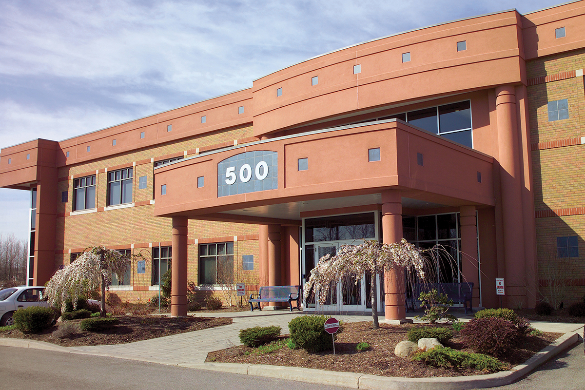
Calkins Corporate Park (Red Creek)
500 Red Creek Drive, Suite 130
Rochester, NY 14623
Lakeside Professional Park
229B Parrish Street, Suite 103
Canandaigua, NY 14424
Jones Memorial Hospital
191 North Main Street
Wellsville, NY 14895
Strong Memorial Hospital
601 Elmwood Avenue
Rochester, NY 14642
Strong West
156 West Avenue
Brockport, NY 14420
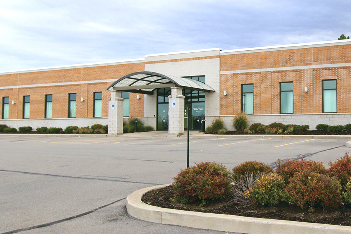
Penfield Crossings Medical Center
2212 Penfield Road, Suite 500
Penfield, NY 14526
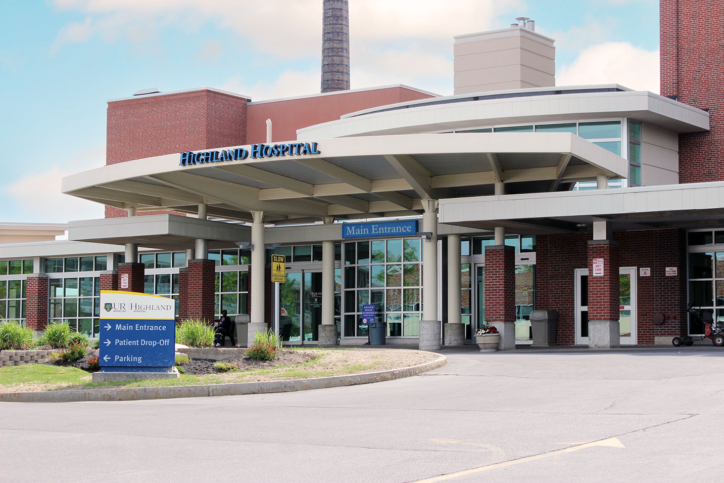
Highland Hospital
1000 South Avenue
Rochester, NY 14620
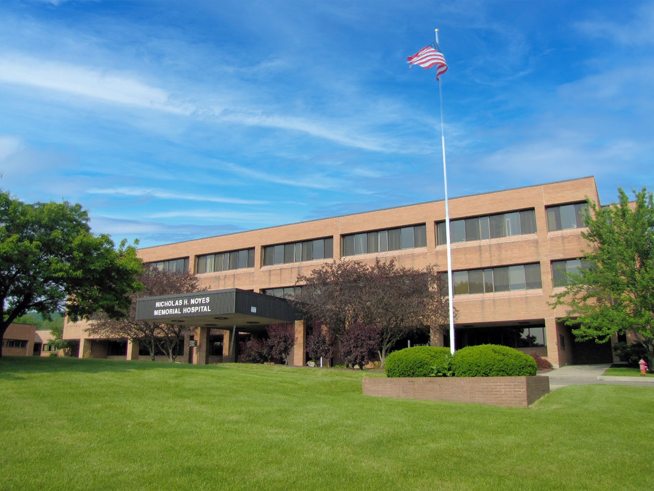
E. Michael Saunder Medical Imaging at Noyes Memorial Hospital
111 Cara Barton Street
Dansville, NY 14437
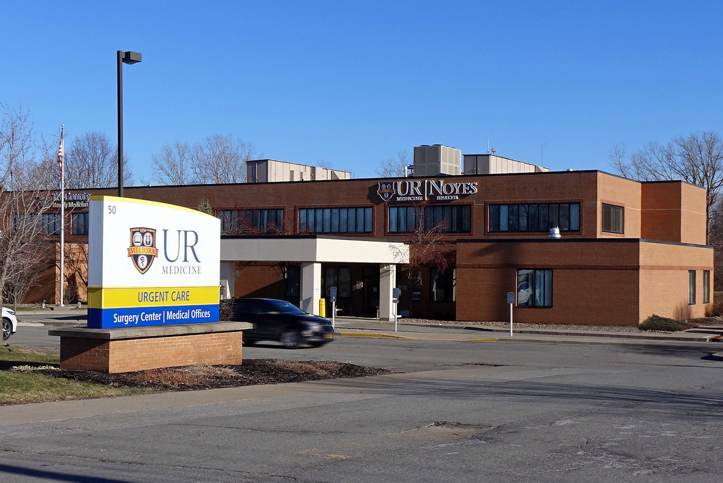
Noyes Health Services
50 East South Street
Geneseo, NY 14454
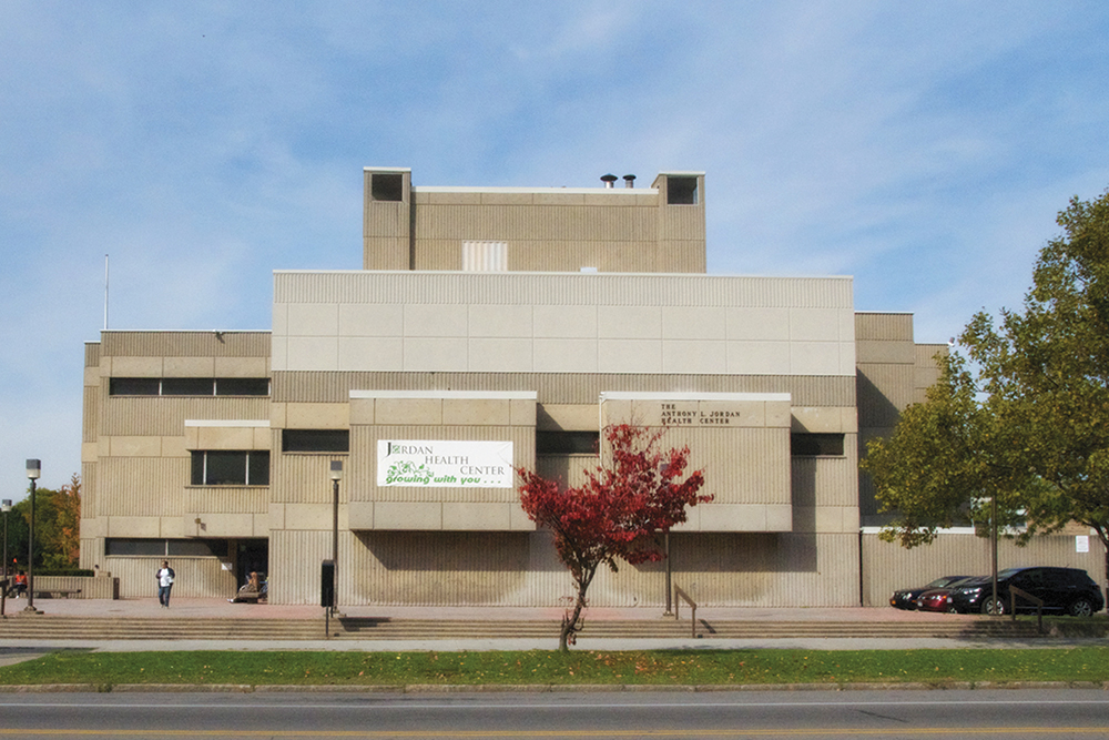
Anthony L. Jordan Health Center
82 Holland Street
Rochester, NY 14605
St. James Medical Office Building
7309 Seneca Road North, Entrance E, Suite 113
Hornell, NY 14843
