Research Projects
Visual Remediation in Schizophrenia
Funded by NIMH. PI, Steven M. Silverstein, PhD.
Many people with schizophrenia experience changes in visual perception. Two well-established changes involve reduction in contrast sensitivity (the ability to detect subtle differences in brightness and shading) and in perceptual organization (the ability to automatically organize the parts of an object or image into a single mental representation). The purpose of this 5-year NIMH-funded project is to determine the effectiveness of three different training programs to improve visual perception in schizophrenia by targeting contrast sensitivity, perceptual organization, or both.
These visual training packages will be compared to changes that occur after a course of standard cognitive remediation. The study involves a comprehensive assessment of different aspects of vision and other functions that depend on vision (e.g., visual memory, reading), and a 30-40 session training program that uses an engaging computer game-like format. Assessment sessions happen before the training; after 20, 30, and 40 sessions; and 6-months after completion of the training program. The program is described in detail in a recent publication that can be accessed here: https://jpbs.hapres.com/htmls/JPBS_1274_Detail.html. The ClinicalTrials.gov registered ID is NCT03314129
Retinal Vasculopathy as a Proxy for Cerebral Vascular Health and a Marker of Cognitive Improvement in Advanced Heart Failure Patients
Funded by the Chernowitz Medical Research Foundation
The purpose of this study is to examine the relationship between blood flow to the brain (using ultrasound of the neck vessels) and cognitive impairment in patients with heart failure. This study recruits patients with heart failure with or without left ventricular assist device (LVAD), patients with diabetic retinopathy, and those without any medical condition. The study also explores the potential role of retinal biomarkers (i.e., the structure and function of the nerve cells and blood vessels in the back of the eye), as predictor of cognition in these patient populations.
Retinal Vasculopathy as a Predictor of Cardiovascular Disease and Risk in Individuals with Early Psychosis
Funded by the URMC Department of Psychiatry Internal Grants Program. PI, Joy Choi, MD.
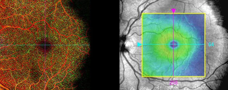
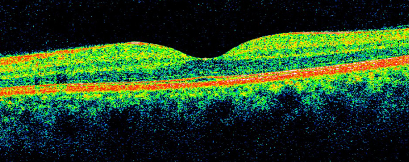
This project examines microvascular changes that impact cardiovascular health in early psychosis with the goal of developing a foundation for primary prevention of cardiovascular disease (CVD) in individuals with psychotic illness. CVD is a major cause of death in individuals with schizophrenia. The exact pathogenesis of CVD in this population is not yet understood but is thought to be multifactorial in origin including genetic predisposition, metabolic sequelae of antipsychotic medications, and unhealthy lifestyles. The inflammatory-vascular model of schizophrenia, supported by the discovery of genes common to both schizophrenia and CVD, hypothesizes that schizophrenia is a neuropsychiatric manifestation of microvascular disease within the central nervous system (CNS) with comorbid systemic microvascular abnormalities. Based on this model, we hypothesize that microvascular changes are present early in the course of schizophrenia in the CNS, and that these correlate with structural and/or functional impairments in the heart. We will use an innovative approach to examining CNS vasculopathy via optical coherence tomography angiography (OCTA), which allows for rapid, high-resolution visualization of the retina – the only part of CNS that can be examined in a non-invasive way. Specifically, we will evaluate in the early psychosis population: 1) abnormality in cardiac structure and function using transthoracic echocardiography, 2) abnormality in retinal vasculature, 3) 10-year CVD risk elevation and 4) the relationship among these measured variables.
Eyeing the Link Between Heart and Brain Health
Funded by the American Heart Association Innovative Project Award. PI, Mark Oldham, MD.
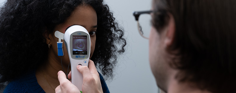
We are exploring the novel application of retinal biomarkers to study delirium risk as well the underlying neural and neurovascular basis for delirium and its subsequent cognitive and functional outcomes. In this study, we are enrolling patients preparing to undergo CABG surgery and evaluating whether the results of retinal exams can identify patients with cognitive impairment before CABG surgery and predict patients with postoperative delirium and cognitive decline. Specifically, we are using retinal optical coherence tomography (OCT), OCT angiography (OCTA) at rest and during vascular reactivity, and flash electroretinography (ERG), which provide direct, efficient, non-invasive measures of neural and neurovascular structure and function in the CNS. The potential application of retinal measures to cognitive health in patients with heart disease could provide a quick and scalable approach to evaluating cognitive vulnerability in this and other populations. Our specific aims are as follows:
Aim 1: To study the association between baseline cognition and ocular biomarkers.
Subjects with lower pre-CABG cognitive performance will have:
H1a. Thinner retinal nerve fiber layer adjacent to optic nerve head, on OCT.
H1b. Reduced retinal perfusion, on OCTA.
H1c. Reduced retinovascular reactivity, on OCTA using a breath-hold maneuver.
H1d. Impaired retinal ganglion cell function, on ERG.
Aim 2: To study baseline ocular biomarkers in relation to post-CABG delirium and severity.
H2a. Post-CABG delirium will be associated with the four retinal indices above.
H2b. Post-CABG delirium severity will be associated with the four retinal indices above.
Aim 3: To explore ocular biomarkers in relation to post-CABG neuropsychiatric outcomes.

Pilot Study Evaluating the Outcomes of a Psychotherapy Protocol for Motivation and Pleasure Symptoms in Clinical High Risk for Psychosis
PI, Tanya Tran, PhD
Dr. Tran is currently conducting a pilot study evaluating the outcomes of a psychotherapy protocol for motivation and pleasure symptoms in individuals with clinical high risk for psychosis. The therapy program adapts a Cognitive Behavioral Therapy framework to augment skills for overcoming cognitive, affective, and behavioral barriers to motivated, goal-directed action in daily life. Secondly, the study will aim to validate a multidimensional battery of assessment tools that are sensitive to changes in negative symptoms within a clinical high risksample and can serve as informative clinical variables to monitor in future, large-scale treatment trials.

A Multicenter, Randomized, Double-Blind, Parallel-Group, Controlled, 16-week Study to Evaluate the Efficacy and Safety of a Digital Therapeutic (CT-155) as an Adjunct to Standard-of-Care Antipsychotic Therapy in Adult and Late Adolescent Participants with Experiential Negative Symptoms of Schizophrenia
PI, Tanya Tran, PhD
Dr. Tran is a local site study coordinator for a multi-site, sponsored trial of a novel digital therapeutic application called CT-155 (developed by Click Therapeutics, Inc. and Boehringer Ingelheim). The study aims to to evaluate the efficacy and safety of CT-155 as an adjunct treatment to standard of care relative to a comparator Digital Control in participants aged 18 years or older diagnosed with experiential negative symptoms of schizophrenia. If demonstrated to be effective, CT-155 may be considered a novel digital therapeutic option as an adjunct treatment for patients diagnosed with experiential negative symptoms of schizophrenia.
Retinal changes associated with psychiatric symptoms
Funded by University of Rochester. PI, Steven M. Silverstein, PhD.
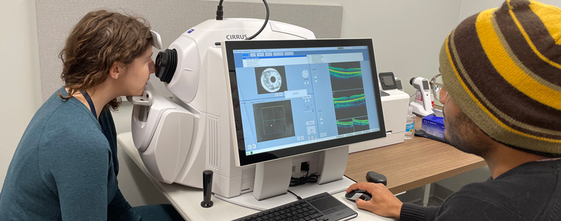
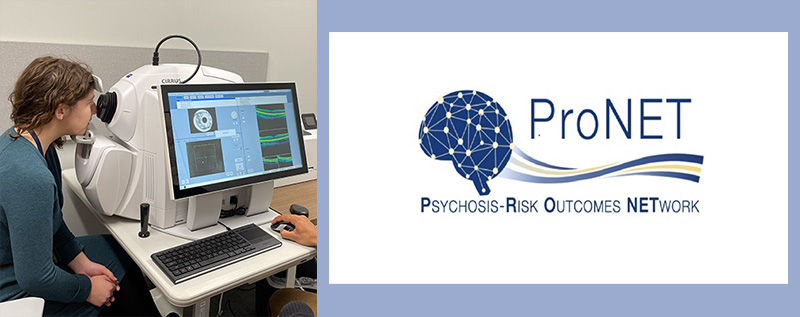
Many studies now demonstrate that changes to the retina (made up of layers of nerve cells and blood vessels, at the back of the eye) are related to changes in the brain and cardiovascular function. These studies are typically in patients with neurological or cardiac disorders, and there are still relatively few studies in psychiatric conditions, although accumulating evidence indicates that psychiatric conditions affect the eye in addition to the brain, and in similar ways. The goal of this study is to determine the kinds of retinal changes that can be observed in people who are being treated for a psychiatric condition. Specifically, we are interested in testing the following groups of people: 1) people with psychotic disorders such as schizophrenia or schizoaffective disorder; 2) people with mood conditions such as bipolar disorder or major depressive disorder; 3) young people (ages 15-28) who are experiencing worsening symptoms over the past year; and 4) people without a psychiatric disorder and without a first degree relative with one of the disorders listed under numbers (1) or (2) above. Participation in the study can be completed in a single day in a session that lasts about 2 hours, including breaks. There are no invasive procedures (e.g., no blood tests, no eye drops, nothing touches the eye, etc.). Retinal imaging is done with short (about 3 second) scans while the participant looks into a tabletop device (see photo below). The rest of the procedures are self-report questionnaires or interviews.
Retinal changes associated with psychiatric symptoms
Funded by University of Rochester. PI, Steven M. Silverstein, PhD
The purpose of this research study is to collect information from individuals who may be at Clinical High Risk (CHR) for possible development of psychosis. People at CHR for psychosis have a higher risk of developing a psychotic disorder than the general population because of particular symptoms and/or family history. These symptoms can include feeling low in mood, feeling paranoid, or seeing or hearing things that they know aren’t there. Some people might worry that their thoughts are being heard, may not feel right in themselves, or might be having more difficulty than usual coping with work, school or relationships. Other people in the CHR group may not experience any of these symptoms, but have a family member with a psychotic disorder which may also increase their risk. For some people, these early symptoms may become more severe over time. For others, the symptoms may stay the same over time, and for others the symptoms may decrease or go away entirely.
Improving our knowledge about symptoms and risk factors may help researchers and clinicians predict possible outcomes for individual participants and develop treatment plans that are suited to the individual participant.Approximately 1430 participants from over 30 study centers worldwide will participate in the ProNET Study. We are interested in young people (ages 12-28), who are noticing a recent change in thinking, behavior, or experiences, such as: a) confusion about what is real or imaginary, b) feeling not in control of their own thoughts of ideas, c) feeling suspicious or paranoid, d) having experiences that may not be real, such as hearing sounds or seeing things that may not be there, e) having trouble communicating clearly. Study description
For more information about the URMC site or to volunteer to participate, please contact Iwona Juskiewicz or call 585-275-4961.
Other studies focused on schizophrenia and/or bipolar disorder that are happening at the University of Rochester Medical Center can be viewed at on the Schizophrenia Treatment and Research Laboratory (STAR) website